Human Reproduction
From gamete formation to the miracle of birth — understanding the anatomy, physiology and hormonal regulation of human reproduction
In this chapter
Humans reproduce sexually and are viviparous — meaning the embryo develops inside the mother's body and is nourished internally. The sequence of reproductive events in humans begins with the formation of gametes: sperms in males and ova (singular: ovum) in females. Once formed, sperms are transferred into the female genital tract during a process called insemination. The fusion of the male and female gametes is known as fertilisation, which gives rise to a zygote.
The zygote then undergoes a series of developmental changes — the blastocyst forms and attaches to the uterine wall (implantation), the embryo develops over roughly nine months (gestation), and finally the baby is delivered (parturition). All of these events occur after the onset of puberty. A key difference between the sexes is that sperm production continues throughout a man's life, whereas ovum formation ceases in women around the age of fifty.
2.1 The Male Reproductive System
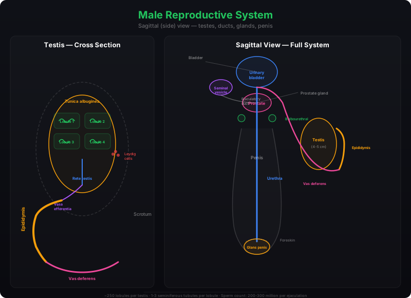
The male reproductive system is located in the pelvis region. It consists of a pair of testes along with accessory ducts, glands, and the external genitalia.
Testes and the Scrotum
The testes are situated outside the abdominal cavity within a pouch called the scrotum. The scrotum plays a vital role in maintaining a temperature approximately 2–2.5 °C lower than the normal internal body temperature — a condition essential for spermatogenesis. In adults, each testis is oval in shape, measuring roughly 4–5 cm in length and 2–3 cm in width. Each testis is enclosed by a dense covering and contains about 250 compartments known as testicular lobules.
Each lobule houses one to three highly coiled seminiferous tubules, the actual sites of sperm production. These tubules are lined internally by two distinct cell types: male germ cells (spermatogonia) that undergo meiosis to form sperm, and Sertoli cells that nourish the developing germ cells.
The regions outside the seminiferous tubules — the interstitial spaces — contain small blood vessels and interstitial cells (also called Leydig cells). Leydig cells are responsible for synthesising and secreting testicular hormones called androgens. Immunologically competent cells are also present in these spaces.
Male Accessory Ducts
The male sex accessory ducts include the rete testis, vasa efferentia, epididymis, and vas deferens. The seminiferous tubules open into the vasa efferentia through the rete testis. The vasa efferentia leave the testis and connect to the epididymis, which lies along the posterior surface of each testis. The epididymis then leads to the vas deferens, which ascends into the abdomen and loops over the urinary bladder. It receives a duct from the seminal vesicle and opens into the urethra as the ejaculatory duct. These ducts collectively store and transport sperms from the testis to the outside through the urethra, which originates from the urinary bladder and extends through the penis.
The Penis
The penis is the male external genitalia. It is composed of special tissue that enables erection, facilitating insemination. The enlarged tip of the penis, called the glans penis, is covered by a loose fold of skin known as the foreskin.
Male Accessory Glands
The male accessory glands include a pair of seminal vesicles, a prostate, and a pair of bulbourethral glands. Their secretions collectively form the seminal plasma, which is rich in fructose, calcium, and certain enzymes. The secretions of the bulbourethral glands also assist in lubricating the penis.
2.2 The Female Reproductive System
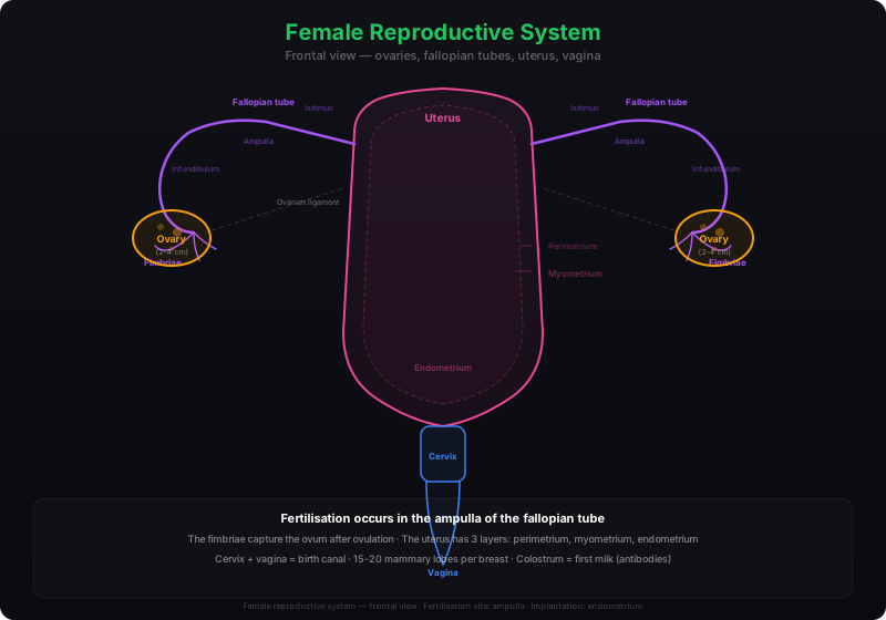
The female reproductive system comprises a pair of ovaries, a pair of oviducts (fallopian tubes), a uterus, a cervix, a vagina, the external genitalia, and a pair of mammary glands. All these parts are structurally and functionally integrated to support ovulation, fertilisation, pregnancy, birth, and child care.
Ovaries
The ovaries are the primary female sex organs. They produce the female gamete (ovum) and several steroid hormones known as ovarian hormones. Each ovary is about 2–4 cm long and is connected to the pelvic wall and uterus by ligaments. A thin epithelium covers each ovary and encloses the ovarian stroma, which is divided into two zones — a peripheral cortex and an inner medulla.
Female Accessory Ducts
The oviducts, uterus, and vagina constitute the female accessory ducts. Each fallopian tube is approximately 10–12 cm long and extends from the periphery of each ovary to the uterus. The part closer to the ovary is a funnel-shaped structure called the infundibulum. The edges of the infundibulum bear finger-like projections called fimbriae, which help capture the ovum after ovulation. The infundibulum opens into a wider region called the ampulla, followed by a narrow-lumen segment called the isthmus, which joins the uterus.
The uterus, also called the womb, is a single, inverted pear-shaped organ supported by ligaments attached to the pelvic wall. It communicates with the vagina through a narrow cervix. The cavity of the cervix, called the cervical canal, along with the vagina, forms the birth canal. The uterine wall consists of three tissue layers:
- Perimetrium — the external thin membranous layer
- Myometrium — the middle thick layer of smooth muscle, responsible for strong contractions during delivery
- Endometrium — the inner glandular layer that lines the uterine cavity and undergoes cyclical changes during the menstrual cycle
External Genitalia
The female external genitalia include the mons pubis, labia majora, labia minora, hymen, and clitoris. The mons pubis is a cushion of fatty tissue covered by skin and pubic hair. The labia majora are fleshy folds that extend downward from the mons pubis and surround the vaginal opening. The labia minora are paired tissue folds lying beneath the labia majora. The vaginal opening is often partially covered by a thin membrane called the hymen. The clitoris is a tiny finger-like structure positioned at the upper junction of the two labia minora, above the urethral opening.
The hymen is frequently torn during the first intercourse, but it can also be broken by a sudden fall, jolt, insertion of a vaginal tampon, or active participation in sports such as horseback riding or cycling. In some women the hymen persists even after coitus. Therefore, the presence or absence of the hymen is not a reliable indicator of virginity or sexual experience.
Mammary Glands
A functional mammary gland is characteristic of all female mammals. These paired structures (breasts) contain glandular tissue and a variable amount of fat. The glandular tissue of each breast is organised into 15–20 mammary lobes, each containing clusters of cells called alveoli. The alveolar cells secrete milk, which is stored in the cavities (lumens) of the alveoli. The alveoli drain into mammary tubules, which from each lobe unite to form a mammary duct. Several mammary ducts merge to form a wider mammary ampulla, which connects to a lactiferous duct and opens at the nipple — the point from which milk is expressed during breastfeeding.
2.3 Gametogenesis
The primary sex organs — the testis in males and the ovaries in females — produce gametes (sperms and ovum, respectively) through a process called gametogenesis.
Spermatogenesis

In the testis, immature male germ cells called spermatogonia reside on the inner wall of the seminiferous tubules. These cells multiply by mitotic division, increasing their numbers. Each spermatogonium is diploid and contains 46 chromosomes. Some spermatogonia, called primary spermatocytes, periodically enter meiosis. A primary spermatocyte completes the first meiotic division (a reduction division), yielding two equal, haploid secondary spermatocytes, each with 23 chromosomes. The secondary spermatocytes then undergo the second meiotic division to produce four equal, haploid spermatids.
The spermatids are subsequently transformed into spermatozoa (sperms) through a process called spermiogenesis. After spermiogenesis, the sperm heads become embedded in the Sertoli cells and are finally released from the seminiferous tubules by a process called spermiation.
Spermatogenesis commences at puberty, triggered by a significant rise in gonadotropin-releasing hormone (GnRH) from the hypothalamus. The elevated GnRH stimulates the anterior pituitary to secrete two gonadotropins: luteinising hormone (LH) and follicle-stimulating hormone (FSH). LH acts on Leydig cells, stimulating androgen synthesis, which in turn promotes spermatogenesis. FSH acts on Sertoli cells, stimulating the secretion of factors that assist in spermiogenesis.
Structure of a Sperm
A sperm is a microscopic structure composed of four distinct parts: a head, a neck, a middle piece, and a tail. A plasma membrane envelops the entire cell. The head contains an elongated haploid nucleus; its anterior portion is capped by a structure called the acrosome, which is filled with enzymes that facilitate fertilisation of the ovum. The middle piece harbours numerous mitochondria that generate the energy required for tail movement — essential for sperm motility and hence for fertilisation.
During a single coitus, the human male ejaculates approximately 200 to 300 million sperms. For normal fertility, at least 60% of the sperms must have normal shape and size, and at least 40% must demonstrate vigorous motility.
Sperms released from the seminiferous tubules are transported by the accessory ducts. Secretions from the epididymis, vas deferens, seminal vesicle, and prostate are essential for sperm maturation and motility. The seminal plasma combined with the sperms constitutes the semen. The functions of the male sex accessory ducts and glands are maintained by testicular hormones (androgens).
Oogenesis
The formation of a mature female gamete is called oogenesis, and it differs markedly from spermatogenesis. Oogenesis is initiated during embryonic development, when several million gamete mother cells (oogonia) form within each fetal ovary. No additional oogonia are produced after birth. These cells begin dividing and enter prophase-I of meiosis, becoming temporarily arrested at this stage as primary oocytes.
Each primary oocyte becomes surrounded by a layer of granulosa cells, forming a primary follicle. A large number of these follicles degenerate between birth and puberty. At puberty, only about 60,000–80,000 primary follicles remain in each ovary. As the primary follicles develop, they acquire additional layers of granulosa cells and a new theca layer, becoming secondary follicles.
The secondary follicle soon transforms into a tertiary follicle, characterised by a fluid-filled cavity called the antrum. The theca layer differentiates into an inner theca interna and an outer theca externa. At this stage, the primary oocyte within the tertiary follicle completes its first meiotic division — an unequal division that produces a large haploid secondary oocyte and a tiny first polar body. The secondary oocyte retains the bulk of the nutrient-rich cytoplasm of the primary oocyte.
The tertiary follicle further matures into the Graafian follicle. The secondary oocyte develops a new membrane around it called the zona pellucida. The Graafian follicle then ruptures, releasing the secondary oocyte (ovum) from the ovary through a process called ovulation.
Comparing Spermatogenesis and Oogenesis
| Feature | Spermatogenesis | Oogenesis |
|---|---|---|
| Site | Seminiferous tubules of testis | Ovaries |
| Onset | At puberty | During embryonic development |
| Division type | Equal cytokinesis throughout | Unequal cytokinesis (polar bodies formed) |
| End products | Four functional spermatids per primary spermatocyte | One functional ovum + polar bodies per primary oocyte |
| Duration | Continuous from puberty onwards | Cyclical; stops at menopause (~50 years) |
| Maturation | Spermiogenesis + spermiation | Completed only upon fertilisation (2nd meiotic division) |
2.4 Menstrual Cycle
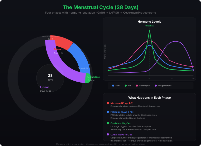
The reproductive cycle observed in female primates — including monkeys, apes, and humans — is called the menstrual cycle. The first occurrence of menstruation, which marks the onset of the cycle, is called menarche. In human females, menstruation repeats at an average interval of about 28–29 days, and the sequence of events spanning from one menstruation to the next constitutes one menstrual cycle. Typically, one ovum is released (ovulation) during the middle of each cycle.
Phases of the Menstrual Cycle
Menstrual Phase: The cycle begins with the menstrual phase, lasting 3–5 days, during which menstrual flow occurs. This flow results from the breakdown of the endometrial lining and its blood vessels, producing a liquid that exits through the vagina. Menstruation occurs only if the released ovum has not been fertilised. A missed period is often the first indication of pregnancy, although other causes such as stress or poor health may also be responsible.
Follicular Phase: Following menstruation, the follicular phase begins. During this phase, the primary follicles in the ovary develop into a fully mature Graafian follicle, while simultaneously the endometrium of the uterus regenerates through proliferation. These changes in the ovary and uterus are driven by shifts in pituitary and ovarian hormone levels. The secretion of gonadotropins (LH and FSH) gradually rises during the follicular phase, stimulating follicular development and the secretion of oestrogens by the growing follicles.
Ovulatory Phase: Both LH and FSH peak around the middle of the cycle (approximately day 14). A rapid surge in LH — the LH surge — triggers the rupture of the Graafian follicle and the release of the ovum (ovulation).
Luteal Phase: After ovulation, the remnants of the Graafian follicle transform into a structure called the corpus luteum. The corpus luteum secretes large quantities of progesterone, which is essential for maintaining the endometrium — a prerequisite for implantation of the fertilised ovum and the subsequent events of pregnancy. If fertilisation does not occur, the corpus luteum degenerates, the endometrium disintegrates, and menstruation begins, marking the start of a new cycle.
During pregnancy, all events of the menstrual cycle cease and menstruation does not occur. In human beings, menstrual cycles typically cease around age 50 — a stage called menopause. Cyclic menstruation is an indicator of a normal reproductive phase, extending from menarche to menopause.
Hormones Regulating the Menstrual Cycle
| Hormone | Source | Role in Menstrual Cycle |
|---|---|---|
| GnRH | Hypothalamus | Stimulates anterior pituitary to release LH and FSH |
| FSH | Anterior pituitary | Promotes follicular growth; stimulates oestrogen secretion by growing follicles |
| LH | Anterior pituitary | LH surge at mid-cycle triggers ovulation; stimulates corpus luteum formation |
| Oestrogens | Growing ovarian follicles | Stimulate endometrial proliferation during the follicular phase |
| Progesterone | Corpus luteum | Maintains the endometrium for implantation; inhibits new follicle development |
2.5 Fertilisation and Implantation
During copulation (coitus), semen is deposited into the vagina by the penis — a process called insemination. The motile sperms swim rapidly through the cervix, enter the uterus, and finally reach the ampullary region of the fallopian tube. The ovum released by the ovary is also transported to the ampullary region, where fertilisation takes place. Since fertilisation demands the simultaneous arrival of both the ovum and sperms to the ampullary region, not every act of copulation results in fertilisation and pregnancy.
The Process of Fertilisation
Fertilisation is the fusion of a sperm with an ovum. When a sperm contacts the zona pellucida layer of the ovum, it triggers membrane changes that block the entry of additional sperms — a phenomenon known as the block to polyspermy. This ensures that only one sperm fertilises a given ovum.
Secretions from the acrosome help the sperm penetrate through the zona pellucida and the plasma membrane of the ovum into its cytoplasm. This entry stimulates the secondary oocyte to complete its second meiotic division — an unequal division that produces a second polar body and a haploid ovum (ootid). The haploid nucleus of the sperm then fuses with the haploid nucleus of the ovum to form a diploid zygote, which contains 46 chromosomes.
The sex of the baby is determined at the moment of fertilisation. Human females have the chromosomal pattern XX, so all ova carry the X chromosome. Human males are XY, meaning approximately 50% of sperms carry an X chromosome and 50% carry a Y chromosome. If an X-bearing sperm fertilises the ovum, the zygote is XX and develops into a female. If a Y-bearing sperm fertilises the ovum, the zygote is XY and develops into a male. Therefore, the sex of the child is determined by the father's sperm, not by the mother.
Cleavage and the Formation of Blastocyst
After fertilisation, the zygote begins to divide mitotically as it moves through the isthmus of the oviduct toward the uterus — a process called cleavage. The division produces 2, 4, 8, and then 16 daughter cells called blastomeres. An embryo with 8 to 16 blastomeres is called a morula.
The morula continues dividing and develops into a blastocyst as it enters the uterus. Within the blastocyst, the blastomeres arrange themselves into two distinct groups: an outer layer called the trophoblast, and an inner cluster of cells called the inner cell mass, which is attached to the trophoblast.
Implantation
The trophoblast layer attaches to the endometrium, while the inner cell mass differentiates into the embryo. After attachment, the uterine cells divide rapidly and envelop the blastocyst. Consequently, the blastocyst becomes embedded in the uterine endometrium — a critical event called implantation, which marks the beginning of pregnancy.
2.6 Pregnancy and Embryonic Development
Formation of the Placenta
Following implantation, finger-like projections called chorionic villi appear on the trophoblast. These villi are surrounded by uterine tissue and maternal blood. The chorionic villi and the uterine tissue interdigitate with each other and together form a structural and functional unit between the developing embryo (foetus) and the maternal body — the placenta.
The placenta facilitates the supply of oxygen and nutrients to the embryo and the removal of carbon dioxide and waste materials produced by the embryo. It is connected to the embryo through the umbilical cord, which enables the transport of substances to and from the embryo. Importantly, the placenta also functions as an endocrine tissue, producing several hormones including:
- hCG (human chorionic gonadotropin)
- hPL (human placental lactogen)
- Oestrogens and progestogens
In the later stages of pregnancy, the ovary also secretes a hormone called relaxin. Notably, hCG, hPL, and relaxin are produced only during pregnancy. Additionally, the levels of several other hormones — including oestrogens, progestogens, cortisol, prolactin, and thyroxine — increase significantly in the maternal blood. This hormonal surge is essential for supporting foetal growth, driving metabolic changes in the mother, and maintaining pregnancy.
Embryonic Development
Immediately after implantation, the inner cell mass (embryo) differentiates into an outer layer called the ectoderm and an inner layer called the endoderm. A third layer, the mesoderm, soon appears between the ectoderm and endoderm. These three germ layers — collectively called the gastrula — give rise to all tissues and organs of the adult body. The inner cell mass contains certain cells called stem cells, which have the potency to differentiate into all tissues and organs.
Milestones of Foetal Development
Human pregnancy lasts approximately nine months. The major milestones during this period are:
End of Month 1
The embryo's heart is formed. The first sign of a growing foetus may be detected by carefully listening to the heartbeat with a stethoscope.
End of Month 2
The foetus develops limbs and digits, marking a recognisable human form.
End of 12 Weeks (1st Trimester)
Most major organ systems are formed. The limbs and external genital organs are well developed.
Month 5
The first movements of the foetus and the appearance of hair on the head are usually observed.
End of 24 Weeks (2nd Trimester)
The body is covered with fine hair, the eyelids separate, and eyelashes are formed.
End of 9 Months
The foetus is fully developed and ready for delivery.
2.7 Parturition and Lactation
Parturition
The average duration of human pregnancy is about nine months, termed the gestation period. At the end of this period, vigorous contractions of the uterus expel the foetus — a process called parturition (childbirth). Parturition is induced by a complex neuroendocrine mechanism.
The signals for parturition originate from the fully developed foetus and the placenta. These signals induce mild uterine contractions called the foetal ejection reflex. This reflex triggers the release of oxytocin from the maternal pituitary. Oxytocin acts on the uterine muscles, causing stronger contractions, which in turn stimulate further oxytocin secretion. This positive feedback loop between uterine contractions and oxytocin release generates progressively stronger contractions, ultimately leading to the expulsion of the baby through the birth canal. Soon after the infant is delivered, the placenta is also expelled from the uterus.
In medical practice, synthetic oxytocin is often injected to induce or strengthen uterine contractions during labour when natural contractions are insufficient.
Lactation
The mammary glands of the female undergo differentiation during pregnancy and begin producing milk towards the end of pregnancy — a process called lactation. This enables the mother to feed the newborn. The milk produced during the first few days of lactation is called colostrum. Colostrum is rich in several antibodies that are absolutely essential for developing resistance in newborn babies. Doctors recommend breastfeeding during the initial period of infant growth to ensure a healthy start for the child.
2.8 Summary
• Humans are sexually reproducing and viviparous organisms. Reproduction involves gametogenesis, insemination, fertilisation, implantation, gestation, and parturition.
• The male reproductive system includes a pair of testes, accessory ducts (rete testis, vasa efferentia, epididymis, vas deferens), glands (seminal vesicles, prostate, bulbourethral glands), and external genitalia (penis). Each testis contains about 250 testicular lobules, each with 1–3 seminiferous tubules.
• The female reproductive system comprises a pair of ovaries, oviducts, uterus, vagina, external genitalia, and mammary glands. The uterus has three layers — perimetrium, myometrium, and endometrium.
• Spermatogenesis begins at puberty under the influence of GnRH, LH, and FSH. A mature sperm has four parts — head, neck, middle piece, and tail. About 200–300 million sperms are ejaculated per coitus.
• Oogenesis begins during embryonic development. Oogonia become primary oocytes, which progress through follicular stages (primary → secondary → tertiary → Graafian follicle) before ovulation releases the secondary oocyte.
• The menstrual cycle has four phases — menstrual, follicular, ovulatory, and luteal — regulated by GnRH, FSH, LH, oestrogens, and progesterone.
• Fertilisation occurs in the ampullary region of the fallopian tube, forming a diploid zygote. The zygote undergoes cleavage to form a morula, then a blastocyst, which implants in the endometrium.
• The placenta develops from chorionic villi and uterine tissue, providing nutrition and gas exchange while secreting hCG, hPL, oestrogens, progestogens, and relaxin.
• Three germ layers — ectoderm, mesoderm, and endoderm — give rise to all adult tissues. Stem cells in the inner cell mass have the potential to form all tissue types.
• Parturition is triggered by the foetal ejection reflex and involves a positive feedback loop of oxytocin secretion and uterine contractions. Colostrum, the first milk, provides essential antibodies to the newborn.