Sexual Reproduction in Flowering Plants
From the development of pollen and embryo sacs to double fertilisation, seed formation, and the fascinating world of apomixis
Table of Contents
1.1 Flower – A Fascinating Organ of Angiosperms
Flowers have captivated human imagination since ancient times. Beyond their aesthetic beauty, colours, scents, and cultural significance, flowers serve a critical biological role: they are the reproductive organs of angiosperms. The myriads of flowers we admire — their colours, fragrances, and intricate structures — exist as aids to sexual reproduction, not merely for human enjoyment.
All flowering plants reproduce sexually. The remarkable diversity of inflorescences, flowers, and floral parts reflects an astonishing range of adaptations aimed at ensuring the formation of fruits and seeds — the end products of sexual reproduction.
To a biologist, a flower is both a morphological marvel and an embryological wonder — the site where sexual reproduction takes place. Flowers are objects of aesthetic, ornamental, social, religious, and cultural value, used as symbols for conveying love, affection, happiness, and grief.
Floriculture — the cultivation of flowers for ornamental purposes — has become a significant industry worldwide. In India alone, major floriculture centres produce roses, marigolds, chrysanthemums, jasmine, and many other species for domestic and export markets.
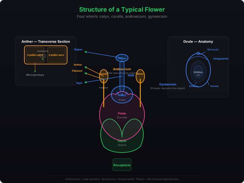
A typical flower has four whorls: the calyx (sepals), corolla (petals), androecium (stamens), and gynoecium (pistils). The two most important units of sexual reproduction develop in the androecium and gynoecium — the male and female reproductive organs, respectively.
1.2 Pre-fertilisation: Structures and Events
Long before a flower appears on a plant, the decision to flower has already been taken. Hormonal and structural changes initiate the differentiation and development of the floral primordium. Inflorescences form, bearing floral buds that eventually open into flowers. Within the flower, the male reproductive organ (androecium) and the female reproductive organ (gynoecium) differentiate and develop.
1.2.1 Stamen, Microsporangium and Pollen Grain
A typical stamen has two parts: a long, slender stalk called the filament, and a terminal, generally bilobed structure called the anther. The proximal end of the filament is attached to the thalamus or the petal of the flower. The number and length of stamens vary greatly across different species — collecting a stamen each from ten different flowers reveals remarkable variation in size and shape.
A typical angiosperm anther is bilobed, with each lobe having two theca — making it dithecous. A longitudinal groove runs lengthwise, separating the theca. In transverse section, the anther appears as a four-sided (tetragonal) structure with four microsporangia located at the corners — two in each lobe.

In transverse section, a typical microsporangium appears nearly circular. It is surrounded by four wall layers:
- Epidermis — outermost protective layer
- Endothecium — helps in dehiscence of the anther
- Middle layers — provide additional protection
- Tapetum — innermost layer; nourishes developing pollen grains; cells have dense cytoplasm and are often multi-nucleate
The outer three layers perform protection and facilitate anther dehiscence to release pollen. The sporogenous tissue occupies the centre of each microsporangium.
Microsporogenesis is the process by which cells of the sporogenous tissue undergo meiotic divisions to form microspore tetrads. Each cell of the sporogenous tissue is a potential pollen or microspore mother cell (PMC). As the anther matures and dehydrates, the microspores dissociate from each other and develop into pollen grains.
Pollen grains represent the male gametophytes. They are generally spherical, measuring about 25–50 micrometres in diameter, with a prominent two-layered wall:
- Exine — hard outer layer made of sporopollenin, one of the most resistant organic materials known; withstands high temperatures, strong acids, and alkali; no known enzyme degrades it
- Intine — thin, continuous inner layer made of cellulose and pectin
The exine has prominent apertures called germ pores where sporopollenin is absent — these are sites where the pollen tube emerges during germination. Pollen grains are well-preserved as fossils because of sporopollenin.
A mature pollen grain contains two cells: a larger vegetative cell (with abundant food reserve and a large irregularly shaped nucleus) and a smaller generative cell (spindle-shaped, floating in the cytoplasm of the vegetative cell). In over 60% of angiosperms, pollen grains are shed at the 2-celled stage. In the remaining species, the generative cell divides mitotically to produce two male gametes before shedding (3-celled stage).
Pollen grains of many species cause severe allergies and bronchial afflictions — asthma, bronchitis, and other chronic respiratory disorders. Parthenium (carrot grass), which entered India as a contaminant with imported wheat, has become ubiquitous and is a major cause of pollen allergy.
Pollen viability is highly variable and depends on temperature and humidity. In cereals like rice and wheat, pollen grains lose viability within 30 minutes of release. In members of Rosaceae, Leguminosae, and Solanaceae, they may remain viable for months. Pollen grains of many species can be stored for years in liquid nitrogen (−196°C) in pollen banks, analogous to seed banks, for crop breeding programmes.
Pollen grains are rich in nutrients and are commercially exploited as food supplements. Pollen tablets and syrups are widely available in western countries and are claimed to enhance the performance of athletes and race horses.
1.2.2 The Pistil, Megasporangium (Ovule) and Embryo Sac
The gynoecium represents the female reproductive part. It may consist of a single pistil (monocarpellary) or more than one (multicarpellary). When multiple pistils are present, they may be fused (syncarpous) or free (apocarpous). Each pistil has three parts: the stigma (landing platform for pollen), the style (elongated slender part), and the ovary (basal bulged part).
Inside the ovary is the ovarian cavity (locule) with the placenta from which the megasporangia — commonly called ovules — arise. The number of ovules per ovary ranges from one (wheat, paddy, mango) to many (papaya, watermelon, orchids).
A typical angiosperm ovule (anatropous) consists of:
- Funicle — stalk attaching the ovule to the placenta
- Hilum — junction between the ovule body and funicle
- Integuments — one or two protective envelopes encircling the nucellus
- Micropyle — small opening at the tip of the integuments
- Chalaza — basal part of the ovule, opposite the micropyle
- Nucellus — mass of cells with abundant reserve food
- Embryo sac (female gametophyte) — located within the nucellus
Megasporogenesis is the process of forming megaspores from the megaspore mother cell (MMC). The MMC differentiates in the micropylar region of the nucellus — it is a large cell with dense cytoplasm and a prominent nucleus. The MMC undergoes meiosis to produce four megaspores.
In most flowering plants, only one megaspore is functional while the other three degenerate. The functional megaspore develops into the female gametophyte (embryo sac). This is called monosporic development.
The nucleus of the functional megaspore divides mitotically to form two nuclei that move to opposite poles (2-nucleate stage). Two more sequential mitotic divisions produce 4-nucleate and then 8-nucleate stages. These divisions are strictly free nuclear — not followed immediately by cell wall formation. After the 8-nucleate stage, cell walls are laid down.
At maturity, the embryo sac is 7-celled and 8-nucleate:
- Egg apparatus (micropylar end) — 2 synergids + 1 egg cell
- Antipodals (chalazal end) — 3 cells
- Central cell — 1 large cell with 2 polar nuclei
The synergids have special cellular thickenings at the micropylar tip called the filiform apparatus, which guides pollen tubes into the synergid.
1.2.3 Pollination
Since male and female gametes in flowering plants are non-motile, they must be brought together for fertilisation. Pollination is the mechanism that achieves this — the transfer of pollen grains from the anther to the stigma of a pistil.
| Type | Description | Key Feature |
|---|---|---|
| Autogamy | Pollination within the same flower | Requires synchrony in pollen release and stigma receptivity; anthers and stigma must lie close together |
| Geitonogamy | Transfer from anther to stigma of another flower on the same plant | Functionally cross-pollination (involves a pollinating agent), but genetically equivalent to autogamy |
| Xenogamy | Transfer from anther to stigma of a different plant | Only type that brings genetically different pollen to the stigma |
Some plants like Viola (common pansy), Oxalis, and Commelina produce two types of flowers: chasmogamous flowers (open, with exposed anthers and stigma) and cleistogamous flowers (never open, with anthers and stigma lying close together). Cleistogamous flowers are invariably autogamous and produce assured seed-set even without pollinators.
Wind Pollination
Pollen grains are light, non-sticky, with well-exposed stamens and large feathery stigmas. Common in grasses and the corn cob. Flowers are not colourful and produce no nectar.
Water Pollination
Rare in flowering plants (~30 genera, mostly monocots). Examples: Vallisneria, Hydrilla, Zostera (seagrass). Pollen grains are often long, ribbon-like, with mucilaginous covering.
Animal Pollination
Most common. Bees, butterflies, flies, beetles, moths, birds, and even bats. Flowers are large, colourful, fragrant, and rich in nectar. Floral rewards include nectar and pollen grains.
Special Relationships
Yucca moth and Yucca plant: neither completes its life cycle without the other. The moth deposits eggs in the ovary; the flower gets pollinated by the moth.
Flowering plants have evolved several mechanisms to discourage self-pollination and encourage cross-pollination, preventing inbreeding depression:
- Unsynchronised timing — pollen released before or after stigma receptivity
- Spatial separation — anther and stigma at different positions
- Self-incompatibility — genetic mechanism inhibiting self-pollen germination or tube growth
- Unisexual flowers — monoecious (castor, maize) prevents autogamy; dioecious (papaya) prevents both autogamy and geitonogamy
Pollen-pistil interaction is a dynamic process. The pistil can recognise compatible pollen (same species) and incompatible pollen (wrong type). Compatible pollen is accepted, and post-pollination events leading to fertilisation proceed. Incompatible pollen is rejected by preventing germination on the stigma or tube growth in the style. This recognition is mediated by chemical components of the pollen interacting with those of the pistil.
Following compatible pollination, the pollen grain germinates on the stigma through a germ pore, producing a pollen tube that grows through the stigma and style to reach the ovary. In 2-celled pollen, the generative cell divides during pollen tube growth; in 3-celled pollen, the tube carries two male gametes from the start. The tube enters the ovule through the micropyle and enters one of the synergids via the filiform apparatus.
You can study pollen germination at home: dust pollen from pea, chickpea, Crotalaria, balsam, or Vinca on a glass slide with a drop of 10% sugar solution. After 15–30 minutes, observe under a low-power microscope — you are likely to see pollen tubes emerging from the grains.
In artificial hybridisation, breeders use emasculation (removing anthers from the flower bud before dehiscence) and bagging (covering with butter paper to prevent contamination). When the stigma becomes receptive, pollen from the desired male parent is dusted on it, and the flower is rebagged. If the female parent has unisexual flowers, emasculation is unnecessary — only bagging is required.
1.3 Double Fertilisation
After the pollen tube enters one of the synergids, it releases two male gametes into the synergid's cytoplasm. Two fusion events then occur simultaneously — making this a process unique to flowering plants.
Syngamy
One male gamete moves towards the egg cell and fuses with its nucleus, completing syngamy and forming a diploid zygote.
Triple Fusion
The other male gamete moves to the two polar nuclei in the central cell and fuses with them, producing a triploid primary endosperm nucleus (PEN). This involves fusion of three haploid nuclei.
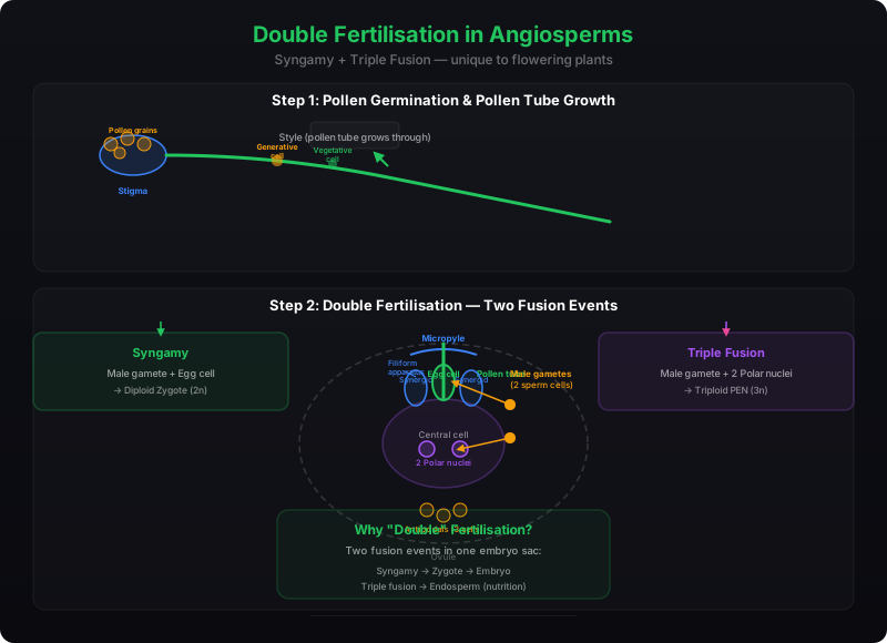
Because two types of fusion — syngamy and triple fusion — take place in each embryo sac, the phenomenon is termed double fertilisation. This is an event unique to angiosperms. The central cell after triple fusion becomes the primary endosperm cell (PEC), which develops into the endosperm, while the zygote develops into the embryo.
1.4 Post-fertilisation: Structures and Events
Following double fertilisation, endosperm and embryo development, maturation of ovules into seeds, and ovary into fruit collectively constitute the post-fertilisation events.
1.4.1 Endosperm
Endosperm development always precedes embryo development — this ensures assured nutrition for the developing embryo. The primary endosperm cell (PEC) divides repeatedly to form a triploid endosperm tissue whose cells are filled with reserve food materials.
In the most common type of development, the PEN undergoes successive nuclear divisions to produce free nuclei — called free-nuclear endosperm. Subsequently, cell wall formation occurs, making the endosperm cellular. The number of free nuclei before cellularisation varies greatly.
The coconut water from a tender coconut is nothing but free-nuclear endosperm (made up of thousands of nuclei), while the surrounding white kernel is the cellular endosperm.
Endosperm may be completely consumed by the developing embryo before seed maturation — as in pea, groundnut, and beans — or it may persist in the mature seed, as in castor and coconut, being used up during germination.
1.4.2 Embryo
The embryo develops at the micropylar end of the embryo sac where the zygote is situated. Most zygotes divide only after a certain amount of endosperm has formed — an adaptation ensuring assured nutrition. The early stages of embryogeny are similar in both monocotyledons and dicotyledons: the zygote produces the proembryo, which progresses through globular, heart-shaped, and mature stages.
Dicot Embryo
Consists of an embryonal axis and two cotyledons. The portion above the cotyledons is the epicotyl (terminating in the plumule). Below is the hypocotyl (terminating in the radicle, covered by root cap).
Monocot Embryo
Has one cotyledon called the scutellum (lateral). The radicle and root cap are enclosed in the coleorrhiza. The epicotyl has a shoot apex and leaf primordia enclosed in the coleoptile.
1.4.3 Seed
The seed is the final product of sexual reproduction in angiosperms — a fertilised ovule formed inside fruits. A seed typically consists of seed coat(s), cotyledon(s), and an embryo axis. The cotyledons are generally thick and swollen due to food storage (as in legumes).
| Seed Type | Description | Examples |
|---|---|---|
| Non-albuminous (ex-albuminous) | No residual endosperm — completely consumed during embryo development | Pea, groundnut, beans |
| Albuminous | Retains part of the endosperm | Wheat, maize, barley, castor |
| With perisperm | Remnants of nucellus persist as perisperm | Black pepper, beet |
As ovules mature into seeds, the integuments harden into tough protective seed coats. The micropyle remains as a small pore facilitating entry of oxygen and water during germination. Mature seeds become relatively dry (10–15% moisture), and the embryo may enter a state of dormancy or germinate if favourable conditions are available.
Simultaneously, the ovary develops into a fruit. The ovary wall becomes the pericarp. Fruits may be fleshy (guava, orange, mango) or dry (groundnut, mustard). In a few species like apple, strawberry, and cashew, the thalamus also contributes to fruit formation — these are called false fruits. Most fruits develop only from the ovary and are called true fruits.
Some species produce fruits without fertilisation — called parthenocarpic fruits. Banana is a familiar example. Parthenocarpy can be induced through growth hormone application, producing seedless fruits.
- Reproductive processes are independent of water — more dependable seed formation
- Better adaptive strategies for dispersal to new habitats
- Sufficient food reserves nourish young seedlings until photosynthesis begins
- Hard seed coat protects the young embryo
- Being products of sexual reproduction, they generate new genetic combinations and variations
Seed viability varies greatly. Some species lose viability within months; others remain viable for centuries. The oldest recorded viable seed is that of Lupinus arcticus, excavated from Arctic Tundra, which germinated after an estimated 10,000 years of dormancy. A 2,000-year-old viable date palm (Phoenix dactylifera) seed was discovered during archaeological excavation at King Herod's palace near the Dead Sea.
1.5 Apomixis and Polyembryony
Although seeds are generally products of fertilisation, some flowering plants — particularly certain species of Asteraceae and grasses — have evolved a mechanism to produce seeds without fertilisation, called apomixis. This is a form of asexual reproduction that mimics sexual reproduction.
There are several ways apomictic seeds develop:
- In some species, the diploid egg cell is formed without reduction division and develops into an embryo without fertilisation
- In many Citrus and Mango varieties, nucellar cells surrounding the embryo sac start dividing, protrude into the embryo sac, and develop into embryos
The occurrence of more than one embryo in a seed is called polyembryony. Orange seeds, for instance, often contain many embryos of different sizes and shapes.
Hybrid varieties of food and vegetable crops have tremendously increased productivity. However, hybrid seeds must be produced every year — if sown, the progeny segregate and lose hybrid characters. Production of hybrid seeds is costly. If hybrids could be made apomictic, there would be no segregation of characters, and farmers could reuse hybrid seeds year after year without purchasing new ones. Active research worldwide aims to understand the genetics of apomixis and transfer apomictic genes into hybrid varieties.
1.6 Summary
• Flowers are the seat of sexual reproduction in angiosperms. The androecium (stamens) represents the male reproductive organs, while the gynoecium (pistils) represents the female reproductive organs.
• A typical anther is bilobed, dithecous, and tetrasporangiate. Pollen grains develop inside microsporangia through microsporogenesis and represent the male gametophytic generation.
• Pollen grains have a two-layered wall — the outer exine (made of sporopollenin, the most resistant organic material) and inner intine (cellulose and pectin). They may be shed at the 2-celled or 3-celled stage.
• The pistil has three parts — stigma, style, and ovary. Ovules within the ovary undergo megasporogenesis to produce the embryo sac (female gametophyte), which is 7-celled and 8-nucleate at maturity.
• Pollination is the transfer of pollen from anther to stigma, mediated by wind, water, or animals. Plants have evolved outbreeding devices — temporal and spatial separation, and self-incompatibility — to promote cross-pollination.
• Pollen-pistil interaction involves chemical dialogue between pollen and pistil, determining compatibility or incompatibility.
• Angiosperms exhibit double fertilisation — syngamy (forming the diploid zygote) and triple fusion (forming the triploid primary endosperm nucleus). This is unique to flowering plants.
• Post-fertilisation events include endosperm development (free-nuclear or cellular), embryo formation, seed maturation (albuminous or non-albuminous), and fruit development (true, false, or parthenocarpic).
• Apomixis is the production of seeds without fertilisation, while polyembryony is the occurrence of multiple embryos in a single seed. Both have agricultural significance.