Anatomy of Flowering Plants
Understanding the internal structure and organisation of tissues, tissue systems, and the anatomy of roots, stems, and leaves in higher plants
In this chapter
In the previous chapter, you studied about the external morphology of plants — their roots, stems, leaves, flowers, fruits, and seeds. But have you ever wondered what lies beneath the surface? How does water travel from the roots all the way up to the tallest leaves of a tree? How are nutrients transported across the plant body?
The answers to these questions lie in the internal structure of plants — a field of study known as anatomy. In this chapter, we will explore how plant bodies are organised at the tissue level, from the meristematic cells that drive growth to the complex permanent tissues that carry out transport, storage, and mechanical support.
A plant body is made up of different kinds of tissues, broadly divided into three major groups: meristematic tissues, permanent tissues, and the tissue systems that these tissues form together. We will also compare the internal anatomy of dicotyledonous and monocotyledonous plants — their roots, stems, and leaves — and conclude with an introduction to secondary growth, the process by which woody plants increase in girth.
6.1 The Tissues
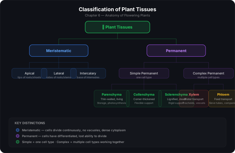
A tissue is a group of cells that are structurally and/or functionally similar and work together to perform a specific role. In plants, tissues are of two fundamental types: meristematic tissues (responsible for growth) and permanent tissues (formed after cells lose the ability to divide and differentiate).
6.1.1 Meristematic Tissues
The growth of plants is largely confined to specialised regions called meristems. These are tissues whose cells are perpetually embryonic — they remain thin-walled, have dense cytoplasm, prominent nuclei, and are capable of continuous cell division. Because of their dividing nature, meristematic cells are typically small, cuboidal, and packed closely together with minimal or no intercellular spaces.
Unlike most plant cells, meristematic cells have a thin cellulosic cell wall, they lack vacuoles (or have very small ones), and they are metabolically very active. Their continuous division ensures that the plant body can grow in length and width throughout its life.
Meristems are classified based on their position in the plant body:
Apical Meristems
Located at the tips of roots and shoots. They produce the primary tissues and are responsible for the primary growth — increase in length of the plant body.
Lateral Meristems
Located along the sides of roots and stems (e.g., vascular cambium, cork cambium). They are responsible for secondary growth — increase in girth or thickness.
Intercalary Meristems
Located at the base of internodes (e.g., in grasses, mint, bamboo). They contribute to elongation of the plant body from the middle regions.
When meristematic cells divide, the outer cells differentiate into permanent tissues while the inner cells continue to divide. This process of a cell taking on a specific structure and function is called differentiation. Cells also undergo division (mitosis), elongation (increase in size), and finally maturation (attaining functional maturity).
6.1.2 Permanent Tissues
Permanent tissues are formed when meristematic cells lose their ability to divide and undergo structural and functional changes. They make up the bulk of the plant body. Permanent tissues are of two types: simple permanent tissues (made of one type of cell) and complex permanent tissues (made of more than one type of cell working together).
Simple Permanent Tissues
These tissues consist of structurally and functionally similar cells. There are three types:
1. Parenchyma
The most common and versatile tissue in plants. Parenchyma consists of relatively unspecialised, thin-walled, living cells that are usually isodiametric (roughly spherical) with intercellular spaces. Their functions include storage of food (starch, oils, water), photosynthesis (when they contain chloroplasts — then called chlorenchyma), and providing buoyancy in aquatic plants (when they contain large air cavities — called aerenchyma).
Chlorenchyma is parenchyma that contains chloroplasts and carries out photosynthesis. Aerenchyma is parenchyma with large air-filled cavities, found in aquatic plants like lotus and water lily, helping them float and facilitating gas exchange.
2. Collenchyma
Collenchyma consists of living cells with unevenly thickened cell walls, particularly at the corners. These cells are elongated and provide flexible mechanical support to the plant body, especially in young stems and petioles. Unlike sclerenchyma, collenchyma cells are alive at maturity and can stretch to accommodate growth — making them ideal for supporting parts of the plant that are still elongating.
Example: The strings you peel from a celery stalk are bundles of collenchyma tissue.
3. Sclerenchyma
Sclerenchyma consists of cells with uniformly thickened, lignified cell walls. These cells are typically dead at maturity and provide rigid mechanical support to the plant body. Sclerenchyma occurs in layers around vascular bundles, in the hard covering of seeds and nuts, and in the husk of coconut.
Sclerenchyma is of two types:
- Fibres — Long, slender, pointed cells found in bundles (e.g., hemp, jute, flax fibres). They are used commercially for making ropes and textiles.
- Sclereids — Short, variable-shaped cells with extremely thick walls. They give the gritty texture to pears and the hardness to seed coats and nut shells.
Complex Permanent Tissues
Complex tissues are made up of more than one type of cell, all working together as a unit. The two major complex tissues in flowering plants are xylem and phloem. Together, they form the vascular tissue of the plant — the transport network that moves water, minerals, and food throughout the body.
Xylem
Xylem is the water-conducting tissue of the plant. It is responsible for the transport of water and dissolved minerals from the roots upward to the stem and leaves. Xylem also provides mechanical support to the plant body. It is composed of four types of elements:
- Tracheids — Long, narrow, dead cells with lignified walls. They conduct water and also provide mechanical support.
- Vessels — Wider, shorter cells arranged end to end, forming continuous pipelines. They are more efficient water conductors than tracheids. Vessels are absent in gymnosperms.
- Xylem parenchyma — Living parenchyma cells that store food and help in lateral conduction of water.
- Xylem fibres — Dead, sclerenchymatous cells that provide mechanical support.
The first-formed xylem is called protoxylem and the later-formed xylem is called metaxylem. In roots, the protoxylem lies towards the periphery and metaxylem towards the centre — this pattern is called exarch. In stems, the arrangement is reversed — called endarch.
Of the four components, tracheids, vessels, and fibres are dead at maturity, while only xylem parenchyma is living. This dead-and-lignified composition is what makes xylem a strong, rigid tissue — it serves as the plant's skeleton.
Phloem
Phloem is the food-conducting tissue. It transports the products of photosynthesis (mainly sucrose) from the leaves to other parts of the plant — roots, fruits, seeds, and growing tips. This process is called translocation. Unlike xylem, phloem transport is an active process requiring energy. Phloem is composed of four types of elements:
- Sieve tubes — Long, tubular cells arranged end to end. They are alive at maturity but lack a nucleus, ribosomes, and most organelles. Their lateral walls have sieve plates with pores for transport.
- Companion cells — Small, specialised parenchyma cells that lie adjacent to sieve tubes. They have a dense cytoplasm and a prominent nucleus, and they regulate the flow of materials in the sieve tubes.
- Phloem parenchyma — Living cells that store food and help in the lateral transport of nutrients.
- Phloem fibres (Bast fibres) — Dead, sclerenchymatous cells that provide mechanical support. Examples include fibres of jute, hemp, and flax.
The first-formed phloem is called protophloem and the later-formed phloem is called metaphloem. Unlike xylem, phloem does not provide major mechanical support since its cells are mostly living and thin-walled.
6.2 The Tissue System
Now that we understand the individual tissues, we can look at how they are organised into tissue systems — functional groupings that span across the plant body. Based on their position, structure, and function, the tissues of flowering plants are classified into three tissue systems:
Epidermal Tissue System
The outermost covering of the plant body. It includes the epidermis, stomata, and epidermal appendages (trichomes and root hairs). It forms a continuous protective layer.
Ground Tissue System
The major portion of the plant body, filling the space between the epidermis and vascular tissues. It includes parenchyma, collenchyma, and sclerenchyma — forming the cortex, pericycle, and pith.
Vascular Tissue System
The transport network of the plant, consisting of xylem and phloem arranged in vascular bundles. It runs through the ground tissue and provides both transport and mechanical support.
6.2.1 Epidermal Tissue System
The epidermal tissue system forms the outermost covering of the entire plant body — roots, stems, leaves, flowers, fruits, and seeds. It comprises:
Epidermis
The epidermis is typically a single layer of compactly arranged, barrel-shaped cells without any intercellular spaces. The outer and lateral walls are often thickened and covered with a waxy layer called the cuticle, which prevents excessive water loss. In xerophytic plants (adapted to dry environments), the cuticle is very thick. In submerged aquatic plants, the cuticle is thin or absent.
In roots, the epidermis bears root hairs — elongated tubular projections that greatly increase the surface area for absorption of water and minerals from the soil.
Stomata
Stomata (singular: stoma) are tiny pores found primarily on the surfaces of leaves, and to a lesser extent on stems and other aerial parts. Each stoma is bounded by a pair of kidney-shaped (in dicots) or dumb-bell-shaped (in monocots) cells called guard cells. Unlike other epidermal cells, guard cells contain chloroplasts.
Stomata regulate gas exchange (CO₂ in, O₂ out) and transpiration (loss of water vapour). When guard cells absorb water and become turgid, the stoma opens; when they lose water and become flaccid, it closes.
In dicot leaves, stomata are more abundant on the lower epidermis (hypostomatic). In monocot leaves (like grasses), stomata are approximately equally distributed on both surfaces (amphistomatic). In floating aquatic plants like Nymphaea, stomata are present only on the upper epidermis.
Epidermal Appendages
The epidermis may bear various appendages:
- Trichomes — Hair-like outgrowths on the epidermis of stems and leaves. They may be glandular (secreting substances) or non-glandular. They protect the plant from herbivory, reduce water loss, and reflect excess light.
- Root hairs — Elongations of epidermal cells in roots, increasing absorption surface area.
6.2.2 Ground Tissue System
The ground tissue system includes all the tissues except the epidermal and vascular tissues. It constitutes the bulk of the plant body and is derived primarily from parenchyma, though it may also contain collenchyma and sclerenchyma.
The ground tissue is divided into three zones:
- Cortex — The region between the epidermis and the vascular cylinder. In stems, the cortex is well-developed and serves for storage and photosynthesis. In roots, it is primarily involved in storage and radial transport.
- Pericycle — The outermost layer of the vascular cylinder (just inside the endodermis). In roots, the pericycle gives rise to lateral roots and is the site where vascular cambium originates during secondary growth.
- Pith (Medulla) — The central region of the stem and root, usually parenchymatous and involved in storage.
In dicot stems, the cortex is clearly differentiated into outer collenchyma, middle parenchyma, and inner endodermis. In monocot stems, the ground tissue is not clearly differentiated into cortex and pith — it appears as a mass of parenchyma surrounding scattered vascular bundles.
6.2.3 Vascular Tissue System
The vascular tissue system consists of xylem and phloem, which are organised into vascular bundles. A vascular bundle typically contains both xylem and phloem together, often with a layer of sclerenchyma called the bundle sheath surrounding them.
Vascular bundles are classified based on the arrangement of xylem and phloem:
- Radial — Xylem and phloem are arranged on different radii, alternating with each other. Found in roots.
- Conjoint — Xylem and phloem are present on the same radius, with phloem towards the outside and xylem towards the inside. Found in stems and leaves.
- Open — Vascular bundles with a strip of vascular cambium between xylem and phloem, capable of secondary growth. Found in dicot stems and roots.
- Closed — Vascular bundles lacking cambium, so no secondary growth is possible. Found in monocot stems and roots.
Table 6.1 — Comparison of Vascular Bundle Types
| Feature | Radial | Conjoint Open | Conjoint Closed |
|---|---|---|---|
| Xylem & Phloem | On different radii | On the same radius | On the same radius |
| Cambium | Absent | Present | Absent |
| Secondary Growth | Not applicable | Occurs | Does not occur |
| Found in | Roots | Dicot stems & roots | Monocot stems |
6.3 Anatomy of Dicotyledonous and Monocotyledonous Plants
Having understood the individual tissues and tissue systems, we can now examine how these are arranged in the roots, stems, and leaves of dicotyledonous and monocotyledonous plants. The differences in their internal anatomy reflect their distinct growth patterns and evolutionary adaptations.
6.3.1 Dicotyledonous Root
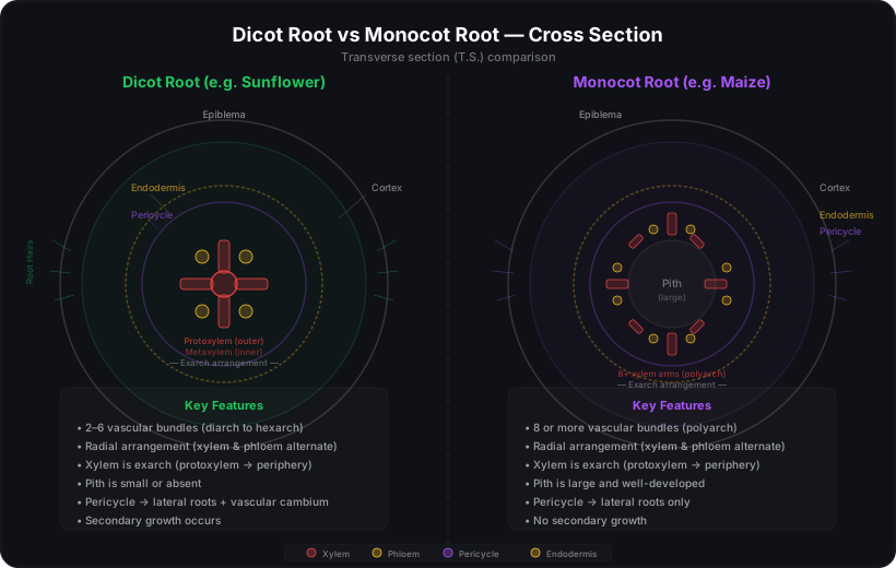
A transverse section (T.S.) of a young dicot root (e.g., sunflower, Helianthus) reveals the following arrangement of tissues, from outside to inside:
- Epiblema — The outermost layer, also called the epidermis or rhizodermis. It bears root hairs and lacks a cuticle.
- Cortex — A wide region of parenchyma cells with intercellular spaces, used for storage of food.
- Endodermis — The innermost layer of the cortex. Its cells have barrel-shaped, thickened radial walls with Casparian strips (bands of suberin) that regulate the passage of water and dissolved substances into the vascular cylinder.
- Pericycle — The layer just inside the endodermis. It is the seat of origin of lateral roots and the vascular cambium during secondary growth.
- Vascular bundles — Typically 2 to 6 (diarch to hexarch) in number. They are radial, with xylem and phloem on alternating radii. Xylem is exarch (protoxylem towards the periphery, metaxylem towards the centre).
- Pith — Small or absent in dicot roots, as the xylem often reaches the centre.
6.3.2 Monocotyledonous Root
A T.S. of a monocot root (e.g., maize, Zea mays) shows a similar general arrangement to the dicot root but with important differences:
- The vascular bundles are more numerous — typically 8 or more (polyarch condition).
- The pith is large and well-developed, unlike the small or absent pith in dicot roots.
- The xylem is also exarch.
- No secondary growth occurs in monocot roots.
Table 6.2 — Dicot Root vs Monocot Root
| Feature | Dicot Root | Monocot Root |
|---|---|---|
| Epiblema | Single-layered, with root hairs | Single-layered, with root hairs |
| Cortex | Widely developed | Widely developed |
| Endodermis | With Casparian strips; barrel-shaped | With Casparian strips; barrel-shaped |
| Pericycle | Gives rise to lateral roots and vascular cambium | Gives rise to lateral roots only |
| Vascular Bundles | 2–6 (diarch to hexarch), radial | 8 or more (polyarch), radial |
| Xylem | Exarch, usually tetrarch | Exarch, polyarch |
| Pith | Small or absent | Large and well-developed |
| Secondary Growth | Occurs | Does not occur |
6.3.3 Dicotyledonous Stem
A T.S. of a young dicot stem (e.g., sunflower) reveals a ring-like arrangement of vascular bundles surrounding a central pith. From outside to inside:
- Epidermis — Single-layered, covered with a thick cuticle. May bear trichomes.
- Cortex — Divided into three zones:
- • Hypodermis — Outermost cortex, made of collenchyma for mechanical support.
- • General cortex — Middle region of parenchyma for storage and photosynthesis.
- • Endodermis — Innermost cortex, also called the "starch sheath" because its cells are rich in starch grains.
- Pericycle — Lies just inside the endodermis. In dicot stems, it consists of sclerenchymatous patches (forming bundle caps above the phloem) and parenchyma.
- Vascular bundles — Arranged in a ring. They are conjoint, open, and endarch (protoxylem towards the centre, metaxylem towards the periphery). Each bundle is capped by sclerenchyma (bundle cap).
- Pith — Large, parenchymatous central region. It may extend as pith rays (medullary rays) between vascular bundles.
The ring-like arrangement of vascular bundles is the hallmark of a dicot stem. This pattern allows the formation of a continuous ring of vascular cambium during secondary growth, enabling the stem to increase in girth.
6.3.4 Monocotyledonous Stem
A T.S. of a monocot stem (e.g., maize) shows a strikingly different arrangement — vascular bundles are scattered throughout the ground tissue, not arranged in a ring. The key features are:
- Epidermis — Single-layered with a thick cuticle. No trichomes typically. Hypodermis is sclerenchymatous (not collenchymatous as in dicot stems).
- Ground tissue — Not differentiated into cortex, pericycle, and pith. It is a continuous mass of parenchyma.
- Vascular bundles — Scattered throughout the ground tissue. They are conjoint, closed (no cambium), and endarch. Each bundle is surrounded by a sclerenchymatous bundle sheath. The bundles are more numerous towards the periphery and fewer towards the centre.
- Pith — Not distinguishable, as the ground tissue is not differentiated.
Two quick diagnostic features:
1. Dicot stem: vascular bundles in a ring, collenchymatous hypodermis, well-differentiated cortex and pith.
2. Monocot stem: vascular bundles scattered, sclerenchymatous hypodermis, no differentiation of ground tissue.
Table 6.3 — Dicot Stem vs Monocot Stem
| Feature | Dicot Stem | Monocot Stem |
|---|---|---|
| Hypodermis | Collenchymatous | Sclerenchymatous |
| Ground Tissue | Differentiated into cortex, pericycle, pith | Not differentiated |
| Vascular Bundles | Ring arrangement | Scattered |
| Bundle Type | Conjoint, open, endarch | Conjoint, closed, endarch |
| Bundle Sheath | Absent or sclerenchymatous caps | Prominent sclerenchymatous sheath |
| Phloem Parenchyma | Present | Absent |
| Secondary Growth | Occurs | Does not occur |
6.3.5 Dorsiventral (Dicotyledonous) Leaf
Dicot leaves are dorsiventral — they have distinct upper (adaxial) and lower (abaxial) surfaces. When a T.S. of a dicot leaf (e.g., mango or mustard) is examined, the following structures are visible:
- Upper epidermis — A single layer of cells covered with a thick cuticle. Stomata are rare or absent on this surface.
- Lower epidermis — Also single-layered, with a thinner cuticle. Stomata are abundant here — this is where most gas exchange and transpiration occurs.
- Mesophyll — The ground tissue between the two epidermal layers. It is differentiated into two types:
- • Palisade parenchyma — Elongated, columnar cells arranged vertically just below the upper epidermis. They are packed with chloroplasts and are the primary site of photosynthesis.
- • Spongy parenchyma — Loosely arranged, oval or rounded cells with large intercellular air spaces below the palisade layer. They facilitate gas exchange.
- Vascular bundles — Running through the mesophyll, forming the leaf veins. Each bundle is surrounded by a bundle sheath of parenchyma or sclerenchyma. The arrangement within is conjoint, closed, and collateral (xylem towards upper side, phloem towards lower side).
6.3.6 Isobilateral (Monocotyledonous) Leaf
Monocot leaves (e.g., maize, grass) are isobilateral — both surfaces are similar in structure and function. Key features of a monocot leaf T.S.:
- Upper and lower epidermis — Both are similar. Stomata are equally distributed on both surfaces (amphistomatic). Bulliform (motor) cells are present in the upper epidermis — they help in rolling and unrolling of the leaf during water stress.
- Mesophyll — Not differentiated into palisade and spongy parenchyma. It consists of a uniform mass of parenchyma with chloroplasts.
- Parallel venation — Veins run parallel to each other. Vascular bundles are arranged in a parallel series.
- Bundle sheath extensions — Sclerenchymatous bundle sheaths may extend to both epidermal surfaces, providing mechanical support.
Table 6.4 — Dicot Leaf vs Monocot Leaf
| Feature | Dicot Leaf (Dorsiventral) | Monocot Leaf (Isobilateral) |
|---|---|---|
| Symmetry | Dorsiventral (different upper & lower surfaces) | Isobilateral (similar both surfaces) |
| Stomata | Mostly on lower epidermis (hypostomatic) | Equally on both surfaces (amphistomatic) |
| Mesophyll | Differentiated into palisade & spongy | Not differentiated |
| Venation | Reticulate | Parallel |
| Bulliform Cells | Absent | Present (in upper epidermis) |
| Palisade Parenchyma | Present (below upper epidermis) | Absent |
The NCERT textbook suggests practical activities:
1. Cut T.S. sections of a young stem from your garden, observe under a microscope, and identify whether it is a monocot or dicot stem based on vascular bundle arrangement.
2. Cut T.S. sections of a dicot and monocot root, and note the differences in the number and arrangement of vascular bundles, size of pith, and presence/absence of cambium.
6.4 Secondary Growth
The growth of roots and stems in length, with the help of apical meristems, is called primary growth. But many plants — especially dicots and gymnosperms — also increase in girth (thickness). This increase in width or thickness is called secondary growth. It is brought about by the activity of two types of lateral meristems: the vascular cambium and the cork cambium (phellogen).
Secondary growth occurs in the roots and stems of dicotyledonous plants and in gymnosperms. It does not occur in monocots — their vascular bundles are closed (lacking cambium), and their ground tissue is not organised into a ring pattern.
6.4.1 Vascular Cambium
The vascular cambium is a thin strip of meristematic cells located between the xylem and phloem. When active, it divides to produce new xylem cells towards the inside and new phloem cells towards the outside, thereby increasing the girth of the stem or root.
Formation in Stems
In a dicot stem, the vascular cambium originates from two sources:
- Intrafascicular cambium — Already present between xylem and phloem in each open vascular bundle.
- Interfascicular cambium — Formed from the medullary rays (pith rays) between the vascular bundles. Cells of the medullary rays become meristematic and join the intrafascicular cambium strips to form a continuous ring of vascular cambium.
Formation in Roots
In dicot roots, the vascular cambium originates primarily from the pericycle — the tissue just outside the phloem. It also develops from parenchyma cells between the xylem and phloem. The cambium first appears as patches opposite the protoxylem poles and eventually forms a complete ring.
Activity of Vascular Cambium
The vascular cambium is variably meristematic — some cells divide while others remain inactive. The cells that divide are called fusiform initials (giving rise to the axial system — tracheary elements and sieve tube elements) and ray initials (giving rise to the radial system — vascular rays).
- Secondary xylem is formed towards the inside of the cambium ring.
- Secondary phloem is formed towards the outside of the cambium ring.
- As the plant ages, more secondary xylem accumulates than secondary phloem. The older secondary phloem gets crushed as the expanding xylem pushes outward.
The accumulated secondary xylem constitutes what we commonly call wood. The lighter-coloured, active secondary xylem formed during spring (with wider vessels) is called spring wood or early wood. The darker, denser secondary xylem formed in autumn (with narrower vessels) is called autumn wood or late wood. The two together form one annual ring, which can be counted to determine the age of a tree.
6.4.2 Cork Cambium (Phellogen)
As the stem grows in girth, the original epidermis ruptures and can no longer protect the plant. A new protective covering called the periderm replaces the epidermis. The periderm is produced by the cork cambium (also called phellogen), a lateral meristem that arises in the cortex region — typically from cells just below the epidermis or in the outer cortex.
- Cork (Phellem) — Produced towards the outside of the cork cambium. Cork cells are dead at maturity, with suberin-coated walls that make them waterproof and resistant to pathogens. They form the outer bark.
- Secondary cortex (Phelloderm) — Produced towards the inside of the cork cambium. These are living parenchymatous cells.
The cork cambium, cork, and secondary cortex together constitute the periderm. The periderm replaces the epidermis as the protective outer covering of the stem.
In woody stems, gas exchange through the impermeable cork layer is facilitated by specialised pores called lenticels. They are lens-shaped openings in the periderm, formed where the cork cambium is active and loose parenchyma cells replace the compact cork cells. Lenticels allow the living tissues beneath the bark to exchange gases with the atmosphere.
6.4.3 Secondary Growth in Dicot Stem — A Complete Picture
Let us now put together the complete sequence of events during secondary growth in a typical dicot stem:

The tissue that lies between the original epidermis and the periderm is called the bark. Bark includes all tissues outside the vascular cambium — secondary phloem, pericycle, cortex, and epidermis (or periderm). Early-formed bark is called primary bark (containing primary phloem), while the rest is secondary bark.
The accumulated secondary xylem (wood) constitutes the bulk of the tree trunk. Heartwood is the dark, hard, central region of old secondary xylem — it is non-functional for transport but provides mechanical support. It is dark due to deposition of resins, tannins, and other substances. Sapwood is the lighter-coloured outer region of secondary xylem that is actively involved in water transport.
Bark is a non-technical term referring to all tissues outside the vascular cambium. This includes secondary phloem, pericycle, cortex, and the periderm (which replaces the epidermis). Cork is just the outermost layer of the periderm — not the entire bark. The commercial cork used for bottle stoppers comes from the cork oak tree (Quercus suber).
6.5 Summary
• The study of the internal structure of plants is called anatomy. Plant tissues are broadly divided into meristematic and permanent tissues.
• Meristematic tissues are found at the tips of roots and shoots (apical), along the sides (lateral), and at the base of internodes (intercalary). They are responsible for growth through cell division.
• Permanent tissues are of two types: simple (parenchyma, collenchyma, sclerenchyma) and complex (xylem and phloem). Xylem conducts water; phloem conducts food.
• Tissues are organised into three tissue systems: epidermal (protective covering), ground (cortex, pericycle, pith), and vascular (xylem and phloem in bundles).
• Dicot and monocot plants differ significantly in their anatomy — in the arrangement of vascular bundles (ring vs scattered), differentiation of ground tissue, and mesophyll of leaves (palisade+spongy vs undifferentiated).
• Secondary growth — increase in girth — occurs in dicots and gymnosperms through the activity of vascular cambium (producing secondary xylem and phloem) and cork cambium (producing periderm that replaces the epidermis).
• The accumulated secondary xylem forms wood, while all tissues outside the vascular cambium constitute the bark. Annual rings can be used to determine the age of a tree.