Biological Classification
How scientists organise the immense diversity of life into systematic groups — from Aristotle to Whittaker's Five Kingdom system
In this chapter
2.1 History of Classification
Since the dawn of civilisation, there have been many attempts to classify living organisms. It was done instinctively — not using criteria that were scientific, but borne out of a need to use organisms for our own use: for food, shelter and clothing.
Aristotle was the earliest to attempt a more scientific basis for classification. He used simple morphological characters to classify plants into trees, shrubs and herbs. He also divided animals into two groups: those which had red blood and those that did not.
In Linnaeus' time, a Two Kingdom system of classification with Plantae and Animalia kingdoms was developed that included all plants and animals respectively. This system did not distinguish between:
- Eukaryotes and prokaryotes
- Unicellular and multicellular organisms
- Photosynthetic (green algae) and non-photosynthetic (fungi) organisms
Classification of organisms into plants and animals was easily done and easy to understand, but a large number of organisms did not fall into either category. Hence the two kingdom classification used for a long time was found inadequate.
Besides gross morphology, a need was felt for including other characteristics like cell structure, nature of wall, mode of nutrition, habitat, methods of reproduction, and evolutionary relationships. Classification systems for living organisms have hence undergone several changes over time.
2.2 Five Kingdom Classification
R.H. Whittaker (1969) proposed a Five Kingdom Classification. The kingdoms defined by him were named Monera, Protista, Fungi, Plantae and Animalia.
The main criteria for classification used by him include: cell structure, body organisation, mode of nutrition, reproduction and phylogenetic relationships.
The three-domain system has also been proposed that divides Kingdom Monera into two domains, leaving the remaining eukaryotic kingdoms in the third domain and thereby a six kingdom classification. You will learn about this system in detail in higher classes.
Table 2.1 — Comparative Account of Five Kingdoms
| Character | Monera | Protista | Fungi | Plantae | Animalia |
|---|---|---|---|---|---|
| Cell type | Prokaryotic | Eukaryotic | Eukaryotic | Eukaryotic | Eukaryotic |
| Cell wall | Non-cellulosic (Polysaccharide + amino acid) | Present in some | Present (chitin) | Present (cellulose) | Absent |
| Nuclear membrane | Absent | Present | Present | Present | Present |
| Body organisation | Cellular | Cellular | Multicellular | Multicellular | Multicellular |
| Mode of nutrition | Autotrophic (chemosynthetic & photosynthetic) and Heterotrophic (saprophytic/parasitic) | Autotrophic (photosynthetic) and Heterotrophic | Heterotrophic (saprophytic/parasitic/symbiotic) | Autotrophic (photosynthetic) | Heterotrophic (holozoic) |
Earlier classification systems included bacteria, blue green algae, fungi, mosses, ferns, gymnosperms and angiosperms under 'Plants'. The character that unified this whole kingdom was that all organisms included had a cell wall in their cells. This placed together groups which widely differed in other characteristics.
When such characteristics were considered, the fungi were placed in a separate kingdom — Kingdom Fungi. All prokaryotic organisms were grouped together under Kingdom Monera and the unicellular eukaryotic organisms were placed in Kingdom Protista.
Kingdom Protista has brought together Chlamydomonas, Chlorella (earlier placed in Algae within Plants, both having cell walls) with Paramecium and Amoeba (which were earlier placed in the animal kingdom, lacking cell wall). This happened because the criteria for classification changed.
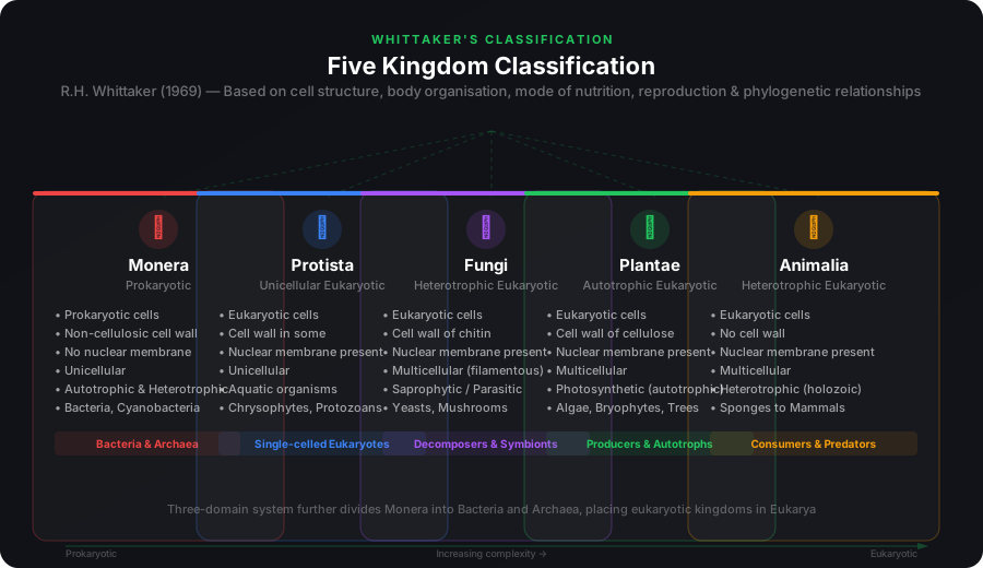
Over time, an attempt has been made to evolve a classification system which reflects not only morphological, physiological and reproductive similarities, but is also phylogenetic — i.e., based on evolutionary relationships.
2.3 Kingdom Monera
Bacteria are the sole members of Kingdom Monera. They are the most abundant micro-organisms. Bacteria occur almost everywhere. Hundreds of bacteria are present in a handful of soil. They also live in extreme habitats such as hot springs, deserts, snow and deep oceans where very few other life forms can survive. Many of them live in or on other organisms as parasites.
Bacterial Shapes
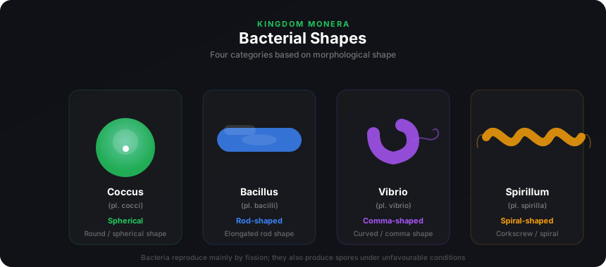
Bacteria are grouped under four categories based on their shape:
Coccus (pl. cocci)
Spherical-shaped bacteria
Bacillus (pl. bacilli)
Rod-shaped bacteria
Vibrium (pl. vibrio)
Comma-shaped bacteria
Spirillum (pl. spirilla)
Spiral-shaped bacteria
Though the bacterial structure is very simple, they are very complex in behaviour. Compared to many other organisms, bacteria as a group show the most extensive metabolic diversity. Some bacteria are autotrophic — they synthesise their own food from inorganic substrates. They may be photosynthetic autotrophic or chemosynthetic autotrophic. The vast majority are heterotrophs.
2.3.1 Archaebacteria
These bacteria are special since they live in some of the most harsh habitats such as extreme salty areas (halophiles), hot springs (thermoacidophiles) and marshy areas (methanogens). Archaebacteria differ from other bacteria in having a different cell wall structure and this feature is responsible for their survival in extreme conditions.
Methanogens are present in the gut of several ruminant animals such as cows and buffaloes and they are responsible for the production of methane (biogas) from the dung of these animals.
2.3.2 Eubacteria
There are thousands of different eubacteria or 'true bacteria'. They are characterised by the presence of a rigid cell wall, and if motile, a flagellum.
The cyanobacteria (also referred to as blue-green algae) have chlorophyll a similar to green plants and are photosynthetic autotrophs. The cyanobacteria are unicellular, colonial or filamentous, freshwater/marine or terrestrial algae. The colonies are generally surrounded by gelatinous sheath. They often form blooms in polluted water bodies.
Some of these organisms can fix atmospheric nitrogen in specialised cells called heterocysts, e.g., Nostoc and Anabaena.
Chemosynthetic autotrophic bacteria oxidise various inorganic substances such as nitrates, nitrites and ammonia and use the released energy for their ATP production. They play a great role in recycling nutrients like nitrogen, phosphorous, iron and sulphur.
Heterotrophic bacteria are most abundant in nature. The majority are important decomposers. Many have significant impact on human affairs — they are helpful in making curd from milk, production of antibiotics, fixing nitrogen in legume roots, etc. Some are pathogens causing diseases like cholera, typhoid, tetanus and citrus canker.
Bacteria reproduce mainly by fission. Sometimes, under unfavourable conditions, they produce spores. They also reproduce by a sort of sexual reproduction by adopting a primitive type of DNA transfer from one bacterium to the other.
Mycoplasma
The Mycoplasma are organisms that completely lack a cell wall. They are the smallest living cells known and can survive without oxygen. Many mycoplasma are pathogenic in animals and plants.
2.4 Kingdom Protista
All single-celled eukaryotes are placed under Protista, but the boundaries of this kingdom are not well defined. What may be 'a photosynthetic protistan' to one biologist may be 'a plant' to another. Members of Protista are primarily aquatic. Being eukaryotes, the protistan cell body contains a well-defined nucleus and other membrane-bound organelles.
2.4.1 Chrysophytes
This group includes diatoms and golden algae (desmids). They are found in fresh water as well as in marine environments. They are microscopic and float passively in water currents (plankton). Most of them are photosynthetic.
In diatoms the cell walls form two thin overlapping shells, which fit together as in a soap box. The walls are embedded with silica and thus the walls are indestructible. Diatoms have left behind large amount of cell wall deposits in their habitat; this accumulation over billions of years is referred to as 'diatomaceous earth'. Being gritty, this soil is used in polishing, filtration of oils and syrups. Diatoms are the chief 'producers' in the oceans.
2.4.2 Dinoflagellates
These organisms are mostly marine and photosynthetic. They appear yellow, green, brown, blue or red depending on the main pigments present in their cells. The cell wall has stiff cellulose plates on the outer surface. Most of them have two flagella — one lies longitudinally and the other transversely in a furrow between the wall plates.
Very often, red dinoflagellates (Example: Gonyaulax) undergo such rapid multiplication that they make the sea appear red — red tides. Toxins released by such large numbers may even kill other marine animals such as fishes.
2.4.3 Euglenoids
Majority are fresh water organisms found in stagnant water. Instead of a cell wall, they have a protein-rich layer called pellicle which makes their body flexible. They have two flagella, a short and a long one. Though they are photosynthetic in the presence of sunlight, when deprived of sunlight they behave like heterotrophs by predating on other smaller organisms. Interestingly, the pigments of euglenoids are identical to those present in higher plants. Example: Euglena.
2.4.4 Slime Moulds
Slime moulds are saprophytic protists. The body moves along decaying twigs and leaves engulfing organic material. Under suitable conditions, they form an aggregation called plasmodium which may grow and spread over several feet. During unfavourable conditions, the plasmodium differentiates and forms fruiting bodies bearing spores at their tips. The spores possess true walls. They are extremely resistant and survive for many years, even under adverse conditions. The spores are dispersed by air currents.
2.4.5 Protozoans
All protozoans are heterotrophs and live as predators or parasites. They are believed to be primitive relatives of animals. There are four major groups:
Amoeboid protozoans: Live in fresh water, sea water or moist soil. They move and capture their prey by putting out pseudopodia (false feet) as in Amoeba. Marine forms have silica shells on their surface. Some such as Entamoeba are parasites.
Flagellated protozoans: Either free-living or parasitic. They have flagella. The parasitic forms cause diseases such as sleeping sickness. Example: Trypanosoma.
Ciliated protozoans: Aquatic, actively moving organisms because of the presence of thousands of cilia. They have a cavity (gullet) that opens to the outside of the cell surface. The coordinated movement of rows of cilia causes the water laden with food to be steered into the gullet. Example: Paramecium.
Sporozoans: Include diverse organisms that have an infectious spore-like stage in their life cycle. The most notorious is Plasmodium (malarial parasite) which causes malaria, a disease which has a staggering effect on human population.
2.5 Kingdom Fungi
The fungi constitute a unique kingdom of heterotrophic organisms. They show a great diversity in morphology and habitat. You must have seen fungi on moist bread and rotten fruits. The common mushroom you eat and toadstools are also fungi. Fungi are cosmopolitan and occur in air, water, soil and on animals and plants. They prefer to grow in warm and humid places.
With the exception of yeasts which are unicellular, fungi are filamentous. Their bodies consist of long, slender thread-like structures called hyphae. The network of hyphae is known as mycelium. Some hyphae are continuous tubes filled with multinucleated cytoplasm — these are called coenocytic hyphae. Others have septae or cross walls in their hyphae. The cell walls of fungi are composed of chitin and polysaccharides.
Most fungi are heterotrophic and absorb soluble organic matter from dead substrates and hence are called saprophytes. Those that depend on living plants and animals are called parasites. They can also live as symbionts — in association with algae as lichens and with roots of higher plants as mycorrhiza.
Reproduction in Fungi
Fungi reproduce sexually by oospores, ascospores and basidiospores. Asexual reproduction is by conidia or sporangiospores or zoospores. The various spores are produced in distinct structures called fruiting bodies.
The sexual cycle involves three steps:
- (i) Plasmogamy — Fusion of protoplasms between two motile or non-motile gametes
- (ii) Karyogamy — Fusion of two nuclei
- (iii) Meiosis — In zygote resulting in haploid spores
In some fungi the fusion of two haploid cells immediately results in diploid cells (2n). However, in other fungi (ascomycetes and basidiomycetes), an intervening dikaryotic stage (n + n, i.e., two nuclei per cell) occurs; such a condition is called a dikaryon and the phase is called dikaryophase of fungus.
Classes of Kingdom Fungi
2.5.1 Phycomycetes
Members are found in aquatic habitats and on decaying wood in moist and damp places or as obligate parasites on plants. The mycelium is aseptate and coenocytic. Asexual reproduction takes place by zoospores (motile) or by aplanospores (non-motile). These spores are endogenously produced in sporangium. A zygospore is formed by fusion of two gametes. Examples: Mucor, Rhizopus (bread mould) and Albugo (parasitic fungi on mustard).
2.5.2 Ascomycetes
Commonly known as sac-fungi, mostly multicellular, e.g., Penicillium, or rarely unicellular, e.g., yeast (Saccharomyces). They are saprophytic, decomposers, parasitic or coprophilous (growing on dung). Mycelium is branched and septate. The asexual spores are conidia produced exogenously on special mycelium called conidiophores. Sexual spores are called ascospores which are produced endogenously in sac-like asci. These asci are arranged in different types of fruiting bodies called ascocarps. Examples: Aspergillus, Claviceps and Neurospora. Many members like morels and truffles are edible and are considered delicacies.
2.5.3 Basidiomycetes
Commonly known forms are mushrooms, bracket fungi or puffballs. They grow in soil, on logs and tree stumps and in living plant bodies as parasites, e.g., rusts and smuts. The mycelium is branched and septate. The asexual spores are generally not found, but vegetative reproduction by fragmentation is common. The sex organs are absent, but plasmogamy is brought about by fusion of two vegetative or somatic cells of different strains or genotypes. The resultant structure is dikaryotic which ultimately gives rise to basidium. Karyogamy and meiosis take place in the basidium producing four basidiospores. The basidiospores are exogenously produced on the basidium. The basidia are arranged in fruiting bodies called basidiocarps. Examples: Agaricus (mushroom), Ustilago (smut) and Puccinia (rust fungus).
2.5.4 Deuteromycetes
Commonly known as imperfect fungi because only the asexual or vegetative phases of these fungi are known. When the sexual forms of these fungi were discovered, they were moved into classes they rightly belong to. The deuteromycetes reproduce only by asexual spores known as conidia. The mycelium is septate and branched. Some members are saprophytes or parasites while a large number of them are decomposers of litter and help in mineral cycling. Examples: Alternaria, Colletotrichum and Trichoderma.
2.6 Kingdom Plantae
Kingdom Plantae includes all eukaryotic chlorophyll-containing organisms commonly called plants. A few members are partially heterotrophic such as the insectivorous plants or parasites. Bladderwort and Venus fly trap are examples of insectivorous plants and Cuscuta is a parasite.
The plant cells have a eukaryotic structure with prominent chloroplasts and cell wall mainly made of cellulose. Plantae includes algae, bryophytes, pteridophytes, gymnosperms and angiosperms.
Life cycle of plants has two distinct phases — the diploid sporophytic and the haploid gametophytic — that alternate with each other. The lengths of the haploid and diploid phases, and whether these phases are free-living or dependent on others, vary among different groups in plants. This phenomenon is called alternation of generations.
2.7 Kingdom Animalia
This kingdom is characterised by heterotrophic eukaryotic organisms that are multicellular and their cells lack cell walls. They directly or indirectly depend on plants for food. They digest their food in an internal cavity and store food reserves as glycogen or fat. Their mode of nutrition is holozoic — by ingestion of food. They follow a definite growth pattern and grow into adults that have a definite shape and size. Higher forms show elaborate sensory and neuromotor mechanism. Most of them are capable of locomotion.
The sexual reproduction is by copulation of male and female followed by embryological development.
2.8 Viruses, Viroids, Prions & Lichens
In the five kingdom classification of Whittaker there is no mention of lichens and some acellular organisms like viruses, viroids and prions.
Viruses
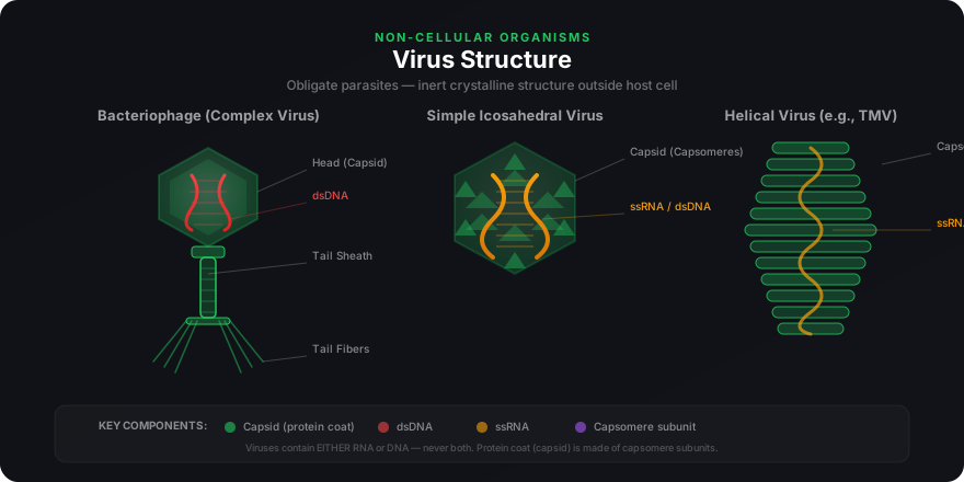
Viruses did not find a place in classification since they are not considered truly 'living', if we understand living as those organisms that have a cell structure. The viruses are non-cellular organisms that are characterised by having an inert crystalline structure outside the living cell. Once they infect a cell, they take over the machinery of the host cell to replicate themselves, killing the host.
Dmitri Ivanowsky (1892) recognised certain microbes as causal organisms of the mosaic disease of tobacco. These were found to be smaller than bacteria because they passed through bacteria-proof filters.
M.W. Beijerinck (1898) demonstrated that the extract of the infected plants of tobacco could cause infection in healthy plants and named the new pathogen "virus" and called the fluid as Contagium vivum fluidum (infectious living fluid).
W.M. Stanley (1935) showed that viruses could be crystallised and crystals consist largely of proteins. They are inert outside their specific host cell. Viruses are obligate parasites.
In addition to proteins, viruses also contain genetic material, that could be either RNA or DNA. No virus contains both RNA and DNA. A virus is a nucleoprotein and the genetic material is infectious. In general, viruses that infect plants have single-stranded RNA and viruses that infect animals have either single or double-stranded RNA or double-stranded DNA. Bacterial viruses or bacteriophages are usually double-stranded DNA viruses.
The protein coat called capsid made of small subunits called capsomeres, protects the nucleic acid. These capsomeres are arranged in helical or polyhedral geometric forms.
Viruses cause diseases like mumps, small pox, herpes and influenza. AIDS in humans is also caused by a virus. In plants, the symptoms can be mosaic formation, leaf rolling and curling, yellowing and vein clearing, dwarfing and stunted growth.
Viroids
In 1971, T.O. Diener discovered a new infectious agent that was smaller than viruses and caused potato spindle tuber disease. It was found to be a free RNA; it lacked the protein coat that is found in viruses, hence the name viroid. The RNA of the viroid was of low molecular weight.
Prions
In modern medicine certain infectious neurological diseases were found to be transmitted by an agent consisting of abnormally folded protein. The agent was similar in size to viruses. These agents were called prions. The most notable diseases caused by prions are bovine spongiform encephalopathy (BSE) commonly called mad cow disease in cattle and its analogous variant Creutzfeldt-Jakob disease (CJD) in humans.
Lichens
Lichens are symbiotic organisms — a mutual association between a fungus (the mycobiont) and an alga (the phycobiont). The fungal partner provides structure, moisture and minerals, while the algal partner provides food through photosynthesis. Lichens are very good pollution indicators — they do not grow in polluted areas.
2.9 Summary
• Biological classification was first proposed by Aristotle on the basis of simple morphological characters. Linnaeus later classified all living organisms into two kingdoms — Plantae and Animalia.
• Whittaker proposed an elaborate five kingdom classification — Monera, Protista, Fungi, Plantae and Animalia. The main criteria were cell structure, body organisation, mode of nutrition and reproduction, and phylogenetic relationships.
• Kingdom Monera includes bacteria — the most abundant micro-organisms showing extensive metabolic diversity. They may be autotrophic or heterotrophic.
• Kingdom Protista includes all single-celled eukaryotes such as Chrysophytes, Dinoflagellates, Euglenoids, Slime-moulds and Protozoans.
• Kingdom Fungi — great diversity in structures and habitat. Most are saprophytic. Phycomycetes, Ascomycetes, Basidiomycetes and Deuteromycetes are the four classes.
• Kingdom Plantae includes all eukaryotic chlorophyll-containing organisms. Life cycle exhibits alternation of generations.
• Kingdom Animalia — heterotrophic eukaryotic, multicellular organisms lacking cell walls. Mode of nutrition is holozoic.
• Viruses are non-cellular, obligate parasites with RNA or DNA. Viroids are free RNA without protein coat. Prions are abnormally folded infectious proteins. Lichens are symbiotic associations useful as pollution indicators.