Locomotion and Movement
How muscles, bones, and joints work together to produce the remarkable movements that define animal life
In this chapter
Movement is one of the most striking features of living organisms. From the streaming of protoplasm in a single-celled Amoeba to the graceful flight of a bird, life is never truly still. Animals and plants exhibit a wide variety of movements — cilia sweeping across a cell surface, flagella propelling a sperm forward, tentacles capturing prey in a Hydra, and limbs lifting us off the ground.
Some of these movements result in a change of place or location. Such voluntary movements are called locomotion. Walking, running, climbing, flying, and swimming are all forms of locomotory movements. Interestingly, the structures used for locomotion may also serve other types of movement. In Paramoecium, for instance, cilia help move food through the cytopharynx as well as propel the organism itself. We use our limbs both to change body posture and to move from one place to another.
The key insight is that all locomotions are movements, but all movements are not locomotions. Locomotion and movement are therefore intimately linked. Animals perform locomotion for various reasons — to search for food, shelter, a mate, suitable breeding grounds, or favourable climatic conditions, or to escape from predators.
17.1 Types of Movement
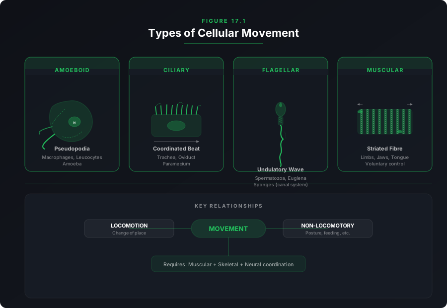
Cells of the human body exhibit three main types of movement: amoeboid, ciliary, and muscular.
Amoeboid Movement
Specialised cells such as macrophages and leucocytes in blood exhibit amoeboid movement. It is effected by pseudopodia formed by the streaming of protoplasm (as seen in Amoeba). Cytoskeletal elements like microfilaments are also involved.
Ciliary Movement
Occurs in most of our internal tubular organs lined by ciliated epithelium. Coordinated ciliary movements in the trachea help remove dust particles and foreign substances. Passage of ova through the female reproductive tract is also facilitated by cilia.
Muscular Movement
Movement of limbs, jaws, tongue, etc., requires muscular movement. The contractile property of muscles is effectively used for locomotion and other movements by human beings and the majority of multicellular organisms.
Flagellar Movement
Flagellar movement helps in the swimming of spermatozoa, maintenance of water current in the canal system of sponges, and locomotion of protists like Euglena. Flagella and cilia are outgrowths of the cell membrane.
Locomotion requires a perfectly coordinated activity of muscular, skeletal, and neural systems. In this chapter, we will explore the types of muscles, their structure, the mechanism of contraction, and important aspects of the skeletal system.
17.2 Muscle
Muscle is a specialised tissue of mesodermal origin. About 40–50 per cent of the body weight of a human adult is contributed by muscles. They possess special properties like excitability, contractility, extensibility, and elasticity.
Muscles have been classified using different criteria — namely, location, appearance, and nature of regulation of their activities. Based on their location, three types of muscles are identified:
Skeletal Muscles
Closely associated with skeletal components of the body. They have a striped appearance under the microscope — hence called striated muscles. Their activities are under voluntary control of the nervous system, so they are also called voluntary muscles. Primarily involved in locomotory actions and changes of body postures.
Visceral Muscles
Located in the inner walls of hollow visceral organs like the alimentary canal and reproductive tract. They do not exhibit any striation and are smooth in appearance — hence called smooth muscles (nonstriated). Their activities are not under voluntary control, making them involuntary muscles. They assist in transporting food through the digestive tract and gametes through the genital tract.
Cardiac Muscles
The muscles of the heart. Many cardiac muscle cells assemble in a branching pattern. Based on appearance, they are striated. They are involuntary in nature — the nervous system does not control their activities directly.
Structure of a Skeletal Muscle
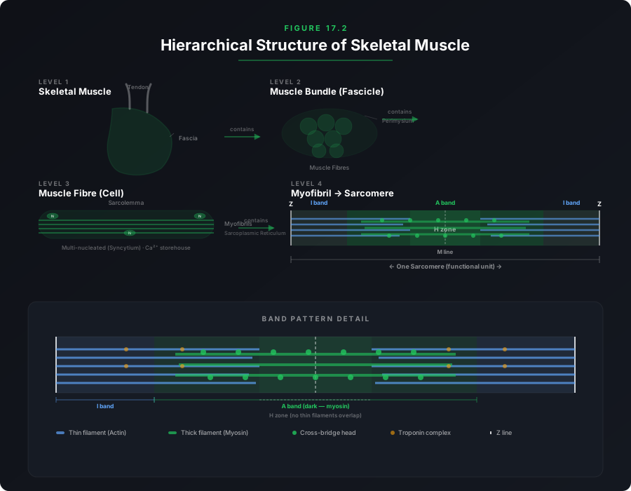
Each organised skeletal muscle in our body is made of a number of muscle bundles (also called fascicles) held together by a common collagenous connective tissue layer called fascia. Each muscle bundle contains a number of muscle fibres (muscle cells).
Each muscle fibre is lined by the plasma membrane called sarcolemma, enclosing the sarcoplasm. Muscle fibre is a syncytium because the sarcoplasm contains many nuclei. The endoplasmic reticulum of the muscle fibres — the sarcoplasmic reticulum — is the storehouse of calcium ions.
A characteristic feature of the muscle fibre is the presence of a large number of parallelly arranged filaments in the sarcoplasm called myofilaments or myofibrils. Each myofibril has alternate dark and light bands on it.
A detailed study of the myofibril has established that the striated appearance is due to the distribution pattern of two important proteins — Actin and Myosin:
- The light bands contain actin and are called I-band (Isotropic band).
- The dark band is called the A-band (Anisotropic band) and contains myosin.
- In the centre of each I-band is an elastic fibre called the Z-line which bisects it. Thin filaments are firmly attached to the Z-line.
- The thick filaments in the A-band are held together in the middle by a thin fibrous membrane called the M-line.
- The portion of the myofibril between two successive Z-lines is considered the functional unit of contraction and is called a sarcomere.
- In a resting state, the edges of thin filaments partially overlap the free ends of the thick filaments, leaving the central part of the thick filaments uncovered. This central part not overlapped by thin filaments is called the H-zone.
17.2.1 Structure of Contractile Proteins
Actin (Thin) Filament
Made of two F-actins (filamentous actins) helically wound to each other. Each F-actin is a polymer of monomeric G-actins (Globular actins). Two filaments of another protein, tropomyosin, also run close to the F-actins throughout its length. A complex protein called troponin is distributed at regular intervals on the tropomyosin. In the resting state, a subunit of troponin masks the active binding sites for myosin on the actin filaments.
Myosin (Thick) Filament
A polymerised protein. Many monomeric proteins called meromyosins constitute one thick filament. Each meromyosin has two important parts: a globular head with a short arm (heavy meromyosin, HMM) and a tail (light meromyosin, LMM). The HMM component — the head and short arm — projects outwards at regular distances and angles from the surface of the polymerised myosin filament and is known as the cross arm. The globular head is an active ATPase enzyme and has binding sites for ATP and active sites for actin.
17.2.2 Mechanism of Muscle Contraction
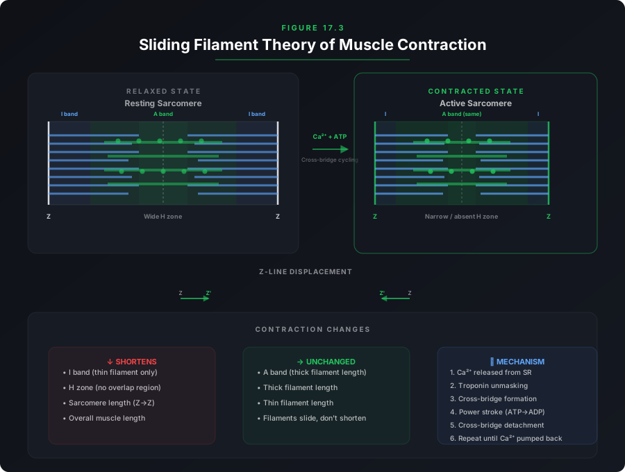
The mechanism of muscle contraction is best explained by the sliding filament theory, which states that contraction of a muscle fibre takes place by the sliding of the thin filaments over the thick filaments.
1. A motor neuron carries a signal to the muscle fibre, generating an action potential in it.
2. This causes the release of Ca²⁺ ions from the sarcoplasmic reticulum into the sarcoplasm.
3. Ca²⁺ ions bind to troponin, causing a conformational change that unmasks the active binding sites on the actin filaments.
4. The myosin heads (with their ATPase activity) bind to the exposed active sites on actin, forming cross bridges.
5. The cross bridges pull the actin filaments, causing them to slide over the myosin filaments. This produces contraction — the Z-lines are pulled closer together, the I-band and H-zone shorten, while the A-band remains unchanged.
6. After the power stroke, ADP and Pi are released. A new ATP molecule binds to the myosin head, causing the cross bridge to break.
7. The myosin head re-cocks (using energy from ATP hydrolysis) and the process repeats, causing further sliding.
8. The process continues until Ca²⁺ ions are pumped back to the sarcoplasmic cisternae, resulting in the re-masking of actin filaments. This causes the Z-lines to return to their original position — relaxation.
Repeated activation of muscles can lead to the accumulation of lactic acid due to anaerobic breakdown of glycogen, causing fatigue.
Red fibres — Contain high amounts of the red-coloured oxygen-storing pigment myoglobin and plenty of mitochondria. They utilise stored oxygen for ATP production and are also called aerobic muscles.
White fibres — Possess very little myoglobin, appear pale or whitish, have few mitochondria, but high amounts of sarcoplasmic reticulum. They depend on anaerobic processes for energy.
17.3 Skeletal System
The skeletal system consists of a framework of bones and a few cartilages. This system has a significant role in movement. Imagine chewing food without jaw bones, or walking without limb bones — both would be impossible!
Bone and cartilage are specialised connective tissues. Bone has a very hard matrix due to calcium salts, while cartilage has a slightly pliable matrix due to chondroitin salts. In human beings, this system is made up of 206 bones and a few cartilages. It is grouped into two principal divisions — the axial and the appendicular skeleton.
Axial Skeleton (80 bones)
The axial skeleton comprises 80 bones distributed along the main axis of the body. The skull, vertebral column, sternum, and ribs constitute the axial skeleton.
🦴 Skull (22 bones)
The skull is composed of two sets of bones — cranial and facial — totalling 22 bones. Cranial bones are 8 in number and form the hard protective outer covering (cranium) for the brain. The facial region is made up of 14 skeletal elements which form the front part of the skull.
A single U-shaped bone called the hyoid is present at the base of the buccal cavity. Each middle ear contains three tiny bones — Malleus, Incus, and Stapes — collectively called ear ossicles. The skull region articulates with the superior region of the vertebral column with the help of two occipital condyles (dicondylic skull).
🦴 Vertebral Column (26 vertebrae)
The vertebral column is formed by 26 serially arranged units called vertebrae and is dorsally placed. It extends from the base of the skull and constitutes the main framework of the trunk. Each vertebra has a central hollow portion (neural canal) through which the spinal cord passes. The first vertebra is the atlas and it articulates with the occipital condyles.
The vertebral column is differentiated into:
| Region | Number | Location |
|---|---|---|
| Cervical | 7 | Neck region (first from skull) |
| Thoracic | 12 | Upper back / chest region |
| Lumbar | 5 | Lower back region |
| Sacral | 1 (fused) | Pelvic region |
| Coccygeal | 1 (fused) | Tail end |
The number of cervical vertebrae are seven in almost all mammals, including human beings. The vertebral column protects the spinal cord, supports the head, and serves as the point of attachment for the ribs and musculature of the back.
🦴 Ribs and Sternum
Sternum is a flat bone on the ventral midline of the thorax. There are 12 pairs of ribs. Each rib is a thin flat bone connected dorsally to the vertebral column and ventrally to the sternum. It has two articulation surfaces on its dorsal end and is hence called bicephalic.
The ribs are classified into three groups:
- True ribs (first 7 pairs) — Attached dorsally to thoracic vertebrae and ventrally connected to the sternum with the help of hyaline cartilage.
- False ribs (vertebrochondral) — 8th, 9th, and 10th pairs — Do not articulate directly with the sternum but join the 7th rib with the help of hyaline cartilage.
- Floating ribs (last 2 pairs — 11th and 12th) — Not connected ventrally and are therefore called floating ribs.
Thoracic vertebrae, ribs, and sternum together form the rib cage.
Appendicular Skeleton
The bones of the limbs along with their girdles constitute the appendicular skeleton. Each limb is made of 30 bones.
Forelimb (Hand) Bones
Humerus, Radius and Ulna, Carpals (wrist bones — 8), Metacarpals (palm bones — 5), and Phalanges (digits — 14).
Hindlimb (Leg) Bones
Femur (thigh bone — the longest bone), Tibia and Fibula, Tarsals (ankle bones — 7), Metatarsals (5), and Phalanges (digits — 14). A cup-shaped bone called Patella covers the knee ventrally (knee cap).
Girdles
🦴 Pectoral Girdle
Each half of the pectoral girdle consists of a clavicle and a scapula. Scapula is a large triangular flat bone situated in the dorsal part of the thorax between the 2nd and 7th ribs. The dorsal, flat, triangular body of scapula has a slightly elevated ridge called the spine, which projects as a flat, expanded process called the acromion. The clavicle articulates with this. Below the acromion is a depression called the glenoid cavity which articulates with the head of the humerus to form the shoulder joint. Each clavicle is a long slender bone with two curvatures — commonly called the collar bone.
🦴 Pelvic Girdle
Consists of two coxal bones. Each coxal bone is formed by the fusion of three bones — ilium, ischium, and pubis. At the point of fusion is a cavity called the acetabulum to which the thigh bone articulates. The two halves of the pelvic girdle meet ventrally to form the pubic symphysis containing fibrous cartilage.
17.4 Joints
Joints are essential for all types of movements involving the bony parts of the body. Joints are points of contact between bones, or between bones and cartilages. Force generated by the muscles is used to carry out movement through joints, where the joint acts as a fulcrum. The movability at these joints varies depending on different factors.
Joints have been classified into three major structural forms:
Fibrous Joints
Do not allow any movement. Shown by the flat skull bones which fuse end-to-end with the help of dense fibrous connective tissues in the form of sutures, to form the cranium.
Cartilaginous Joints
The bones involved are joined together with the help of cartilages. The joint between adjacent vertebrae in the vertebral column is of this pattern and it permits limited movements.
Synovial Joints
Characterised by the presence of a fluid-filled synovial cavity between the articulating surfaces of the two bones. This arrangement allows considerable movement. These joints help in locomotion and many other movements.
Types of Synovial Joints
| Type of Joint | Location | Example |
|---|---|---|
| Ball and Socket | Between humerus and pectoral girdle | Shoulder joint |
| Hinge | Knee joint | Knee |
| Pivot | Between atlas and axis | Neck rotation |
| Gliding | Between the carpals | Wrist movement |
| Saddle | Between carpal and metacarpal of thumb | Thumb opposition |
17.5 Disorders of Muscular and Skeletal System
Several disorders affect the muscular and skeletal systems. Understanding these conditions is essential for appreciating the importance of a healthy locomotory system.
Myasthenia Gravis
An autoimmune disorder affecting the neuromuscular junction, leading to fatigue, weakening, and paralysis of skeletal muscle.
Muscular Dystrophy
Progressive degeneration of skeletal muscle, mostly due to a genetic disorder.
Tetany
Rapid spasms (wild contractions) in muscle due to low Ca²⁺ in body fluid.
Arthritis
Inflammation of joints. Can be caused by wear and tear, autoimmune conditions, or infection.
Osteoporosis
Age-related disorder characterised by decreased bone mass and increased chances of fractures. Decreased levels of estrogen is a common cause.
Gout
Inflammation of joints due to accumulation of uric acid crystals.
17.6 Summary
• Movement is an essential feature of all living beings. A voluntary movement that causes an animal to change its place is called locomotion. Animals move generally in search of food, shelter, mate, breeding ground, better climate, or to protect themselves.
• The cells of the human body exhibit amoeboid, ciliary, and muscular movements. Locomotion and many other movements require coordinated muscular activities.
• Three types of muscles are present in our body: Skeletal muscles (striated, voluntary), Visceral muscles (nonstriated, involuntary), and Cardiac muscles (striated, branched, involuntary). Muscles possess excitability, contractility, extensibility, and elasticity.
• Muscle fibre is the anatomical unit of muscle. Each muscle fibre has many parallelly arranged myofibrils. Each myofibril contains many serially arranged units called sarcomeres — the functional units of contraction. Each sarcomere has a central A band made of thick myosin filaments, and two half I bands made of thin actin filaments on either side, marked by Z lines.
• Actin and myosin are polymerised proteins with contractility. The active sites for myosin on resting actin filament are masked by troponin. Myosin head contains ATPase and has ATP binding sites and active sites for actin.
• A motor neuron carries signal to the muscle fibre which generates an action potential. This causes the release of Ca²⁺ from sarcoplasmic reticulum. Ca²⁺ activates actin which binds to the myosin head to form cross bridges. These cross bridges pull the actin filaments causing them to slide over the myosin filaments — contraction. Ca²⁺ are then returned to sarcoplasmic reticulum, inactivating actin. Cross bridges break and the muscles relax.
• Repeated stimulation leads to fatigue. Muscles are classified as Red fibres (high myoglobin, aerobic) and White fibres (low myoglobin, anaerobic).
• Bones and cartilages constitute the skeletal system. The skeletal system is divisible into axial (skull, vertebral column, ribs, sternum — 80 bones) and appendicular (limb bones and girdles — 126 bones).
• Three types of joints: fibrous (no movement), cartilaginous (limited movement), and synovial (considerable movement). Synovial joints play a significant role in locomotion.