Body Fluids and Circulation
Understanding the composition of blood and lymph, the human heart, cardiac cycle, double circulation, and disorders of the circulatory system
Table of Contents
15.1 Blood
Every living cell in our body needs a constant supply of nutrients, oxygen, and other essential substances. At the same time, waste and harmful by-products of metabolism must be removed continuously to keep tissues healthy. Different organisms have evolved different strategies to achieve this. Simple animals like sponges and coelenterates simply circulate water from their surroundings through their body cavities. More complex organisms, however, rely on specialised internal fluids to transport these substances.
In most higher animals — including humans — the primary transport medium is blood. A second body fluid, lymph, also plays an important supporting role. Blood is a special type of connective tissue consisting of a fluid matrix called plasma and several types of formed elements suspended within it.
Blood is classified as a connective tissue because it consists of cells (formed elements) embedded in a fluid extracellular matrix (plasma), just as cartilage or bone has cells in a matrix. It is unique among connective tissues because its matrix is liquid.
15.1.1 Plasma
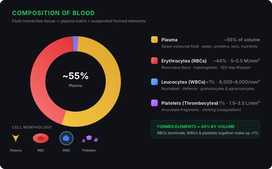
Plasma is the straw-coloured, slightly viscous fluid portion of blood. It makes up approximately 55 per cent of total blood volume. About 90–92 per cent of plasma is water, while proteins account for 6–8 per cent.
The three major groups of plasma proteins are:
- Fibrinogen — Essential for blood clotting (coagulation). When activated, it is converted into fibrin threads that form the structural basis of a clot.
- Globulins — Primarily involved in the body's defence mechanisms. They include antibodies that help fight infections.
- Albumins — Help maintain osmotic balance between blood and surrounding tissues. They account for the largest fraction of plasma proteins.
In addition to proteins, plasma also contains small amounts of minerals such as Na⁺, Ca²⁺, Mg²⁺, HCO₃⁻, Cl⁻, and other ions. It carries glucose, amino acids, lipids, and other nutrients that are always in transit through the body. Coagulation factors are also present in plasma in an inactive form. When all clotting factors are removed from plasma, the remaining fluid is called serum.
15.1.2 Formed Elements
The cellular components of blood — erythrocytes, leucocytes, and platelets — are collectively referred to as formed elements. They make up about 45 per cent of blood volume.
Erythrocytes (Red Blood Cells)
Erythrocytes, or red blood cells (RBCs), are the most numerous cells in blood. A healthy adult male has approximately 5 to 5.5 million RBCs per mm³ of blood. RBCs are formed in the red bone marrow in adults. In most mammals, mature RBCs lack a nucleus and have a biconcave disc shape, which increases the surface area for gas exchange.
These cells contain an iron-bearing, red-coloured protein called haemoglobin, which gives blood its characteristic colour and is critical for transporting respiratory gases — oxygen and carbon dioxide. A healthy individual has about 12–16 grams of haemoglobin per 100 mL of blood. RBCs have an average lifespan of about 120 days, after which they are destroyed in the spleen, often referred to as the "graveyard of RBCs."
Leucocytes (White Blood Cells)
Leucocytes, or white blood cells (WBCs), are colourless because they lack haemoglobin. Unlike RBCs, they are nucleated and are relatively fewer in number — typically 6,000 to 8,000 per mm³ of blood. WBCs are generally short-lived and are the body's primary defence against pathogens.
WBCs are classified into two main categories based on the presence or absence of granules in their cytoplasm:
Granulocytes
Neutrophils (60–65%): Most abundant; phagocytic cells that engulf invading bacteria.
Eosinophils (2–3%): Associated with allergic reactions and parasitic infections.
Basophils (0.5–1%): Secrete histamine, serotonin, and heparin; involved in inflammatory responses.
Agranulocytes
Lymphocytes (20–25%): Responsible for immune responses. Two major types — B-lymphocytes and T-lymphocytes.
Monocytes (6–8%): Largest WBCs; phagocytic cells that destroy foreign organisms entering the body.
Platelets (Thrombocytes)
Platelets, also called thrombocytes, are small, colourless, anucleate cell fragments produced from large bone marrow cells called megakaryocytes. A normal count is 1,50,000 to 3,50,000 platelets per mm³ of blood. When a blood vessel is injured, platelets release substances that trigger coagulation (blood clotting), preventing excessive blood loss. A significant drop in platelet count can lead to bleeding disorders.
All formed elements of blood originate from a common precursor — the haemocytoblast, a stem cell in the red bone marrow. Depending on the chemical signals they receive, haemocytoblasts differentiate into erythrocytes, various types of leucocytes, or megakaryocytes (which give rise to platelets).
15.1.3 Blood Groups
Human blood differs from person to person in certain immunological characteristics, even though it looks similar. Two major systems of blood grouping are widely used worldwide: the ABO system and the Rh system.
15.1.3.1 ABO Grouping
ABO grouping is based on the presence or absence of two surface antigens — designated A and B — on the surface of RBCs. These antigens are chemicals that can trigger an immune response. Similarly, the plasma of different individuals contains two natural antibodies: anti-A and anti-B, which are produced in response to antigens that are not present on the individual's own RBCs.
| Blood Group | Antigens on RBCs | Antibodies in Plasma | Can Donate To |
|---|---|---|---|
| A | A | Anti-B | A, AB |
| B | B | Anti-A | B, AB |
| AB | A, B | None | AB only |
| O | None | Anti-A, Anti-B | A, B, AB, O |
During blood transfusion, donor and recipient blood must be carefully matched to avoid agglutination (clumping/destruction of RBCs). Individuals with blood group O are called universal donors because their RBCs lack both A and B antigens and can be safely given to anyone. Individuals with blood group AB are called universal recipients because their plasma contains no anti-A or anti-B antibodies and can therefore receive blood from any group.
15.1.3.2 Rh Grouping
A separate antigen known as the Rh factor (named after the Rhesus monkey in which it was first discovered) is also found on the surface of RBCs in the majority — roughly 80 per cent — of humans. People who possess this antigen are designated Rh⁺ve (Rh positive), while those who lack it are Rh⁻ve (Rh negative).
An Rh⁻ve individual, if exposed to Rh⁺ve blood, will produce specific anti-Rh antibodies. Therefore, Rh compatibility must also be checked before transfusions.
Erythroblastosis foetalis: A special case arises when an Rh⁻ve mother carries an Rh⁺ve foetus. During the first pregnancy, the two blood circulations are separated by the placenta, so the mother's immune system does not encounter Rh antigens. However, during delivery, small amounts of the foetus's Rh⁺ve blood may mix with the mother's blood, causing her to produce anti-Rh antibodies. In any subsequent pregnancies with an Rh⁺ve foetus, these maternal antibodies can cross the placenta, attack the foetal RBCs, and cause severe anaemia or jaundice — a condition called erythroblastosis foetalis. This can be prevented by administering anti-Rh antibodies (RhoGAM) to the Rh⁻ve mother immediately after the delivery of the first child.
15.1.4 Coagulation of Blood
When you cut your finger, bleeding does not continue indefinitely — the blood eventually stops flowing. This is due to coagulation or blood clotting, a protective mechanism that prevents excessive blood loss from the body.
The process works as follows: The injured site develops a dark reddish-brown mass called a coagulum (clot), which is essentially a network of fine protein threads called fibrins. Within this mesh, dead and damaged blood cells become trapped.
Fibrins are formed from an inactive precursor in the plasma called fibrinogen. The enzyme responsible for this conversion is thrombin. Thrombin itself is produced from another inactive plasma protein called prothrombin. An enzyme complex known as thrombokinase (or thromboplastin) catalyses the conversion of prothrombin to thrombin. Thrombokinase is generated through a series of linked enzymatic reactions — a process called the clotting cascade.
The clotting cascade involves a sequence of clotting factors, each activating the next in the chain. This amplification system ensures that even a small initial trigger can produce a rapid, large-scale clotting response. Calcium ions (Ca²⁺) play an essential role at several steps in this cascade.
15.2 Lymph (Tissue Fluid)
As blood flows through the capillaries in body tissues, some water along with small water-soluble substances leak out into the spaces between the cells. The larger proteins and most of the formed elements, however, remain within the blood vessels. This leaked fluid is called interstitial fluid or tissue fluid.
Tissue fluid has essentially the same mineral composition as plasma. All exchange of nutrients, gases, and wastes between the blood and the cells takes place through this fluid.
An extensive network of thin-walled vessels called the lymphatic system collects this interstitial fluid and drains it back into the major veins. The fluid present within this lymphatic system is called lymph. Lymph is a colourless fluid that contains specialised lymphocytes responsible for the body's immune responses. It also serves as an important carrier for nutrients and hormones.
Fats are absorbed not directly into the blood but through the lymphatic vessels. Specialised lymphatic capillaries called lacteals, present in the finger-like projections of the small intestine (intestinal villi), absorb dietary fats and transport them as part of the lymph.
15.3 Circulatory Pathways
Animals display two fundamental patterns of circulation:
Open Circulatory System
Found in arthropods and molluscs. Blood (called haemolymph) is pumped by the heart into large vessels that empty into open body cavities called sinuses. Here, the blood directly bathes the organs before slowly draining back to the heart.
Closed Circulatory System
Found in annelids and chordates (including all vertebrates). Blood is always contained within a continuous network of blood vessels. This allows more precise regulation of blood flow and pressure.
All vertebrates possess a muscular, chambered heart. The number of chambers varies across groups:
| Vertebrate Group | Heart Chambers | Circulation Type |
|---|---|---|
| Fishes | 2 chambers (1 atrium, 1 ventricle) | Single circulation |
| Amphibians & Reptiles (except crocodiles) | 3 chambers (2 atria, 1 ventricle) | Incomplete double circulation |
| Crocodiles, Birds & Mammals | 4 chambers (2 atria, 2 ventricles) | Complete double circulation |
In fishes, the heart pumps deoxygenated blood to the gills, where it gets oxygenated. This oxygen-rich blood then flows to the body tissues. Deoxygenated blood from the tissues returns to the heart. Because blood passes through the heart only once in a complete circuit, this is called single circulation.
In amphibians and reptiles, the left atrium receives oxygenated blood from the lungs/skin, while the right atrium receives deoxygenated blood from the body. However, because there is only one ventricle, some mixing of oxygenated and deoxygenated blood occurs. This is termed incomplete double circulation.
In birds and mammals, the separation is complete. Oxygenated and deoxygenated blood received by the left and right atria respectively flows into separate ventricles. The ventricles pump the blood into appropriate circuits without mixing — this is complete double circulation.
15.3.1 Human Circulatory System
The human circulatory system — also called the blood vascular system — consists of a muscular, chambered heart, a network of closed, branching blood vessels, and blood.
The Heart
The heart is a mesodermally derived organ located in the thoracic cavity between the two lungs, slightly tilted towards the left. It is roughly the size of a clenched fist. The heart is protected by a double-walled, membranous sac called the pericardium, which encloses the pericardial fluid that cushions and lubricates the heart.
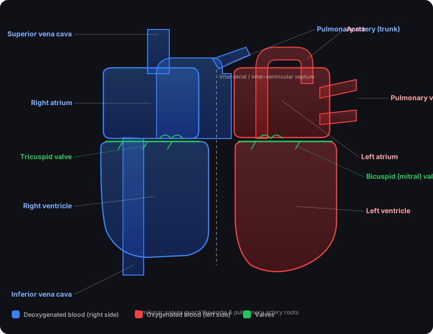
The human heart has four chambers: two relatively smaller, upper chambers called atria (singular: atrium) and two larger, lower chambers called ventricles. A thin muscular wall called the inter-atrial septum separates the right and left atria, while a thicker wall, the inter-ventricular septum, separates the two ventricles.
The atrium and ventricle on the same side are separated by a thick fibrous tissue called the atrio-ventricular septum, but each septum has an opening that allows the two chambers to communicate:
- Right side: The opening between the right atrium and right ventricle is guarded by the tricuspid valve, composed of three muscular flaps (cusps).
- Left side: The opening between the left atrium and left ventricle is guarded by the bicuspid (mitral) valve.
The openings of the right and left ventricles into the pulmonary artery and the aorta, respectively, are guarded by semilunar valves. These valves allow blood to flow in only one direction — from atria to ventricles, and from ventricles to the arteries — and prevent any backward flow.
Nodal Tissue
The entire heart is made of cardiac muscle, and the ventricular walls are much thicker than those of the atria. A specialised type of cardiac muscle called nodal tissue is distributed at specific locations in the heart:
- Sino-atrial node (SAN): A patch of nodal tissue located in the upper right corner of the right atrium. It can generate the maximum number of action potentials — about 70–75 per minute — and is therefore called the pacemaker of the heart.
- Atrio-ventricular node (AVN): Another mass of nodal tissue located in the lower left corner of the right atrium, close to the atrio-ventricular septum.
- Atrio-ventricular bundle (AV bundle): A bundle of nodal fibres that extends from the AVN, passes through the atrio-ventricular septum, emerges on top of the inter-ventricular septum, and immediately divides into right and left bundles.
- Purkinje fibres: Minute fibres that extend throughout the ventricular musculature on both sides, arising from the bundle branches. They rapidly transmit the impulse so that both ventricles contract nearly simultaneously.
Nodal tissue has the intrinsic ability to generate action potentials without any external stimulus — it is autoexcitable. However, different parts of the nodal system generate action potentials at different rates. The SAN generates the most (70–75 per minute), making it the natural pacemaker. Our heart normally beats about 70–75 times per minute (average 72 beats per minute).
15.3.2 Cardiac Cycle
The heart functions through a precisely coordinated sequence of events called the cardiac cycle. Each cycle lasts about 0.8 seconds and consists of the following phases:
Joint Diastole (Relaxation Phase)
Initially, all four chambers of the heart are in a relaxed state. The tricuspid and bicuspid valves are open, so blood from the pulmonary veins and vena cava flows into the left and right ventricles, respectively, through the atria. The semilunar valves are closed at this stage.
Atrial Systole (Atrial Contraction)
The SAN generates an action potential that stimulates both atria to contract simultaneously. This atrial systole pushes an additional volume of blood into the ventricles, increasing ventricular filling by about 30 per cent.
Ventricular Systole (Ventricular Conduction & Contraction)
The action potential is conducted from the SAN to the ventricular side via the AVN and AV bundle. The AVN generates an action potential that stimulates a contraction of the ventricles. The ventricular pressure rises, causing the bicuspid and tricuspid valves to close — this produces the first heart sound, "lub."
As ventricular pressure continues to increase, the semilunar valves guarding the pulmonary artery and the aorta are forced open, allowing blood to flow into the arterial circuits.
Ventricular Diastole (Relaxation)
The ventricles relax, and ventricular pressure falls. This causes the semilunar valves to close — producing the second heart sound, "dub" — preventing backflow of blood into the ventricles. As pressure continues to decline, the tricuspid and bicuspid valves open again under the pressure of blood accumulated in the atria from the veins. Blood flows freely into the ventricles once more, and the cycle begins again.
Heart rate: ~72 beats per minute (average)
Duration of one cardiac cycle: 0.8 seconds
Stroke volume: ~70 mL per ventricle per beat
Cardiac output: ~5 litres per minute (5000 mL/min) for a healthy individual
Cardiac output = Stroke volume × Heart rate
The body can alter both stroke volume and heart rate to adjust cardiac output — for example, athletes have a significantly higher cardiac output than sedentary individuals.
Heart Sounds
During each cardiac cycle, two distinct sounds can be heard through a stethoscope:
- First heart sound ("lub"): Produced by the closure of the tricuspid and bicuspid (atrio-ventricular) valves at the start of ventricular systole.
- Second heart sound ("dub"): Produced by the closure of the semilunar valves at the start of ventricular diastole.
These sounds are of great clinical significance because any deviation from the normal pattern can indicate a heart valve disorder or other cardiac abnormality.
15.3.3 Electrocardiogram (ECG)
The electrical activity of the heart can be recorded from the body surface using a device called an electrocardiograph. The resulting record is called an electrocardiogram (ECG). In a standard ECG, the patient is connected to the machine with three leads — one attached to each wrist and one to the left ankle — that continuously monitor the heart's electrical activity.
Each peak in the ECG waveform is labelled with a letter from P to T, each corresponding to a specific electrical event:
- P-wave: Represents the depolarisation (electrical excitation) of the atria, which leads to atrial contraction.
- QRS complex: Represents the depolarisation of the ventricles, initiating ventricular contraction. The contraction begins shortly after Q and marks the onset of systole.
- T-wave: Represents the repolarisation (return to normal state) of the ventricles. The end of the T-wave marks the end of systole.
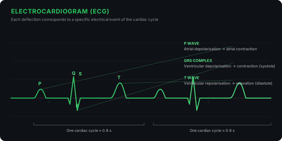
By counting the number of QRS complexes in a given time period, one can determine the heart rate. Since ECGs obtained from different individuals show roughly the same shape for a given lead configuration, any deviation from this pattern indicates a possible abnormality or disease — making the ECG an invaluable diagnostic tool in cardiology.
15.4 Double Circulation
Blood flows through a fixed route of blood vessels — arteries and veins. Each type of vessel has a characteristic wall structure with three layers:
- Tunica intima — the innermost lining of squamous endothelium
- Tunica media — a middle layer of smooth muscle and elastic fibres
- Tunica externa — an outermost layer of fibrous connective tissue with collagen fibres
The tunica media is relatively thinner in veins than in arteries, which is why veins are more compliant and have a lower blood pressure than arteries.
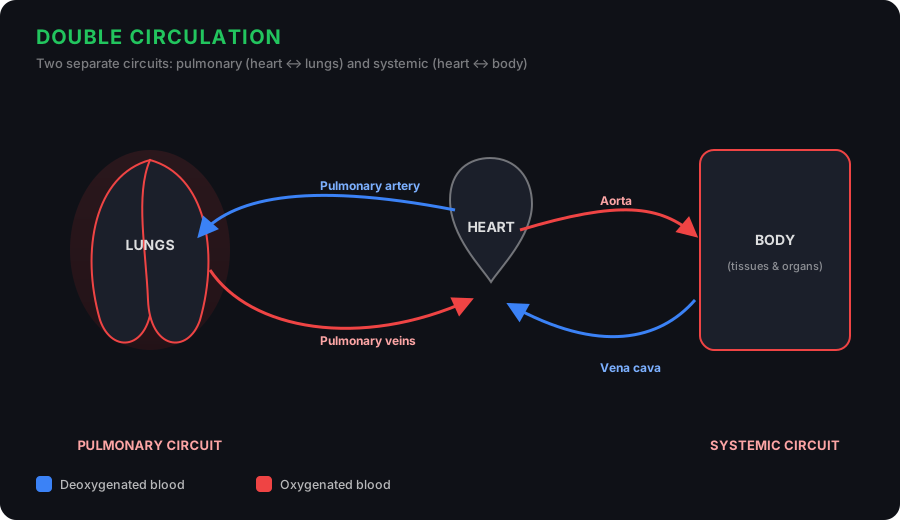
In humans, the circulatory system has two distinct but interconnected pathways:
Pulmonary Circulation
Deoxygenated blood is pumped by the right ventricle into the pulmonary artery, which carries it to the lungs. In the lung capillaries, blood picks up oxygen and releases carbon dioxide. The oxygenated blood then returns to the left atrium via the pulmonary veins.
Systemic Circulation
Oxygenated blood entering the left atrium is pumped by the left ventricle into the aorta. A network of arteries, arterioles, and capillaries delivers it to all body tissues. Deoxygenated blood is collected by venules, veins, and the vena cava, and returned to the right atrium.
Systemic circulation delivers oxygen and nutrients to every tissue and carries away carbon dioxide and wastes. This pathway provides an opportunity for the blood to be fully oxygenated before being pumped to the body — a critical advantage for maintaining the high metabolic rate of warm-blooded animals. The pulmonary circulation ensures that deoxygenated blood is continually refreshed at the lungs.
Hepatic Portal System
A special arrangement called the hepatic portal system connects the digestive system to the liver. The hepatic portal vein carries nutrient-rich blood from the intestines to the liver before it enters the general systemic circulation. This allows the liver to process, detoxify, and store nutrients before they reach the rest of the body.
In addition, a dedicated coronary circulatory system supplies blood to and from the heart muscle (cardiac musculature) itself, ensuring that the heart — which never rests — always receives the oxygen and nutrients it needs.
15.5 Regulation of Cardiac Activity
The normal activities of the heart are regulated intrinsically — that is, by the specialised nodal tissue within the heart itself. This property is called autorhythmicity, and because of it, the heart is said to be myogenic.
However, cardiac function can also be modulated by neural and hormonal mechanisms:
- Sympathetic nervous system: Neural signals through sympathetic nerves (part of the autonomic nervous system) can increase the heart rate, strengthen ventricular contraction, and thereby raise cardiac output. Adrenal medullary hormones can also increase cardiac output.
- Parasympathetic nervous system: Parasympathetic neural signals (via the vagus nerve) decrease the heart rate, slow the speed of action potential conduction, and thereby reduce cardiac output.
A special neural centre in the medulla oblongata of the brain integrates signals from both the sympathetic and parasympathetic branches to maintain an optimal heart rate. This autonomic regulation ensures that cardiac output can be quickly adjusted in response to changing demands — such as during exercise, stress, or rest.
15.6 Disorders of Circulatory System
Several common and serious conditions can affect the circulatory system. Here are the major ones:
High Blood Pressure (Hypertension)
Normal blood pressure is about 120/80 mm Hg, where 120 is the systolic (pumping) pressure and 80 is the diastolic (resting) pressure. If repeated measurements read 140/90 or higher, it indicates hypertension. Persistent high blood pressure damages blood vessels and can lead to heart disease, stroke, and kidney damage.
Coronary Artery Disease (CAD)
Also known as atherosclerosis, CAD affects the arteries supplying blood to the heart muscle. Deposits of calcium, fat, cholesterol, and fibrous tissue accumulate on the inner walls, narrowing the arterial lumen and restricting blood flow to the heart.
Angina (Angina Pectoris)
Characterised by acute chest pain that occurs when the heart muscle does not receive enough oxygen. It is more common among middle-aged and elderly individuals and arises from conditions that reduce blood flow to the heart.
Heart Failure
A condition in which the heart cannot pump blood effectively enough to meet the body's needs. Also called congestive heart failure because fluid congestion in the lungs is a primary symptom. Importantly, heart failure is distinct from cardiac arrest (the heart stops beating entirely) and heart attack (sudden damage to heart muscle from inadequate blood supply).
✦ Chapter Summary
- Blood is a fluid connective tissue that transports nutrients, oxygen, and waste substances to and from cells. Lymph (tissue fluid) assists in this transport for certain substances.
- Blood consists of a fluid matrix (plasma) and formed elements: RBCs (erythrocytes), WBCs (leucocytes), and platelets (thrombocytes).
- Human blood groups — A, B, AB, and O — are based on A and B surface antigens on RBCs. The Rh factor provides another basis for grouping.
- Tissue fluid, derived from blood, fills the spaces between cells. Lymph is essentially tissue fluid that has entered the lymphatic system and is similar to blood except in protein content and formed elements.
- All vertebrates and some invertebrates have a closed circulatory system. The human system consists of a four-chambered heart, a network of blood vessels, and blood.
- The sino-atrial node (SAN) is the pacemaker, generating 70–75 action potentials per minute.
- The cardiac cycle lasts 0.8 seconds and involves the sequential contraction and relaxation of atria and ventricles.
- ECG is the electrical recording of the heart's activity and is of great clinical significance.
- Humans have a complete double circulation: pulmonary (lungs) and systemic (body) circuits.
- Though the heart is autoexcitable, its activity is modulated by neural and hormonal mechanisms.